
10.1 Hypersensitivity #
10.1.1 Four types #
Gell and Coombs sorted damaging responses by effector. Types I–III are antibody and type IV is T cells: a normal mechanism of earlier chapters aimed at a harmless antigen or at the host.
| Type | Effector | Timing | Mechanism | Example |
|---|---|---|---|---|
| I | IgE on mast cells | Minutes | Mediator release after cross-linking | Anaphylaxis |
| II | IgG or IgM on a surface | Hours | Complement, phagocytosis, ADCC, or altered signalling | Haemolytic disease of the newborn; drug-induced haemolysis |
| III | Soluble immune complexes | Hours locally; about one to two weeks in serum sickness | Deposition, complement, neutrophils | Serum sickness |
| IV | T cells | About 48–72 hours | Macrophage activation or killing | Tuberculin skin test |
10.1.2 Type I #
A first exposure in a person biased to Th2 switches B cells to IgE, which occupies FcεRI. Re-exposure cross-links it. Within minutes, histamine and leukotrienes cause leak, bronchoconstriction, mucus, and itch; over the next hours, IL-5 and TNF bring eosinophils. Hay fever, asthma, and urticaria are local forms. Anaphylaxis is the same mechanism in several organs at once: generalised leak drops the blood pressure, and bronchial smooth muscle constricts.
10.1.3 Types II and III #
Type II antibody binds a fixed cell or matrix antigen, and complement, phagocytes, or ADCC damage it, or the antibody changes a receptor’s behaviour. In haemolytic disease of the newborn, an RhD-negative mother sensitised by an earlier pregnancy makes IgG against RhD. IgG crosses the placenta on FcRn. Her own cells lack the antigen; foetal cells that carry it are destroyed. Penicillin can act as a hapten on the red-cell surface and produce the same clearance. In Graves disease, IgG stimulates the TSH receptor and the thyroid over-produces hormone. Type III antibody binds a soluble antigen. Large complexes are carried on red-cell CR1 to liver and spleen. Intermediate complexes deposit in vessels, joints, and glomeruli, fix complement, and bring neutrophils. Serum sickness is the prototype: antibody appears while foreign serum antigen is still present, and about one to two weeks later there is fever, urticaria, joint pain, and proteinuria. Lupus injures tissue by this effector.
10.1.4 Type IV #
Type IV is delayed because the effector is a memory T cell, not pre-formed antibody. In the tuberculin skin test, mycobacterial protein recalls memory Th1 cells; IFN-γ and macrophages produce a palpable induration at about two to three days. A positive test reports memory. It does not by itself report active disease or protection. Contact hypersensitivity to a hapten such as nickel or urushiol is the same type, and CD8 cells can kill haptenated keratinocytes. Serum does not transfer these reactions.
10.2 Autoimmunity #
10.2.1 How tolerance fails #
Autoimmunity is escape of particular clones from the tolerance of Chapters 6 and 8. Only single-gene disasters such as IPEX and AIRE deficiency are global. Established routes, which can combine, are failure to display tissue peptides in the thymus, failure of Foxp3 regulation, and molecular mimicry. Rheumatic fever after streptococcal infection, with antibody that binds both M protein and heart, is the classical mimicry example. Apoptotic nuclei that C1q fails to clear can immunise. HLA type, which decides which peptides are presented, is the strongest common genetic association. Identical twins are not fully concordant, so environment contributes, often without one established trigger.
10.2.2 One organ, and the whole body #
Type 1 diabetes is organ-specific. CD8 and CD4 cells destroy insulin-producing β cells. Antibodies to insulin, GAD65, IA-2, and ZnT8 mark the process more than they cause it, and clinical diabetes appears after most β cells are already lost. HLA-DR3 and HLA-DR4 raise the risk. Graves disease is also organ-specific, but the effector is stimulating antibody, type II, and the gland is overactive rather than destroyed. Hashimoto thyroiditis is the destructive T-cell disease of the same organ. Systemic lupus erythematosus is systemic because the antigens — double-stranded DNA, histones, ribonucleoproteins such as Sm — are in every cell. Anti-dsDNA tracks renal disease and activity; anti-Sm is more specific. Damage is largely type III, with consumption of C3 and C4. Nucleic acid in those complexes engages TLR7 and TLR9, driving type I interferon and further B-cell activation. The disease predominates in women of childbearing age.
10.3 Immunodeficiency #
10.3.1 Primary defects that illuminate the pathway #
Severe combined immunodeficiency is adaptive failure from infancy. X-linked loss of the common γ chain removes IL-7 signalling, so T cells do not develop, and removes IL-15, so NK cells do not; B cells are present and functionally poor (T-negative, B-positive, NK-negative). RAG1 or RAG2 deficiency prevents every antigen-receptor rearrangement, so T and B cells are absent and NK cells, which do not use RAG, are spared. Adenosine deaminase deficiency poisons all three developing lineages. DiGeorge syndrome is the anatomical T-cell version: the thymus fails with the third and fourth pharyngeal pouches. X-linked agammaglobulinaemia stops B cells only. Bruton tyrosine kinase signals from the pre-B-cell receptor; development halts there, mature B cells are almost absent, and every isotype is profoundly low, while T cells are intact. Boys present after maternal IgG wanes, around the middle of the first year, with pyogenic infection. CD40-ligand deficiency (X-linked hyper-IgM syndrome) leaves IgM and blocks switching, germinal centres, and CD40-dependent macrophage activation, so cell-mediated infections appear as well. Chronic granulomatous disease removes the oxidase. Terminal complement deficiency removes the pore and presents as recurrent invasive Neisseria. C3 deficiency removes opsonisation from every route. C1q deficiency presents as lupus-like disease. MHC class II deficiency, from loss of CIITA or an RFX factor, leaves almost no CD4 cells and resembles SCID. MHC class I deficiency, from loss of TAP or tapasin, reduces CD8 cells and is milder.
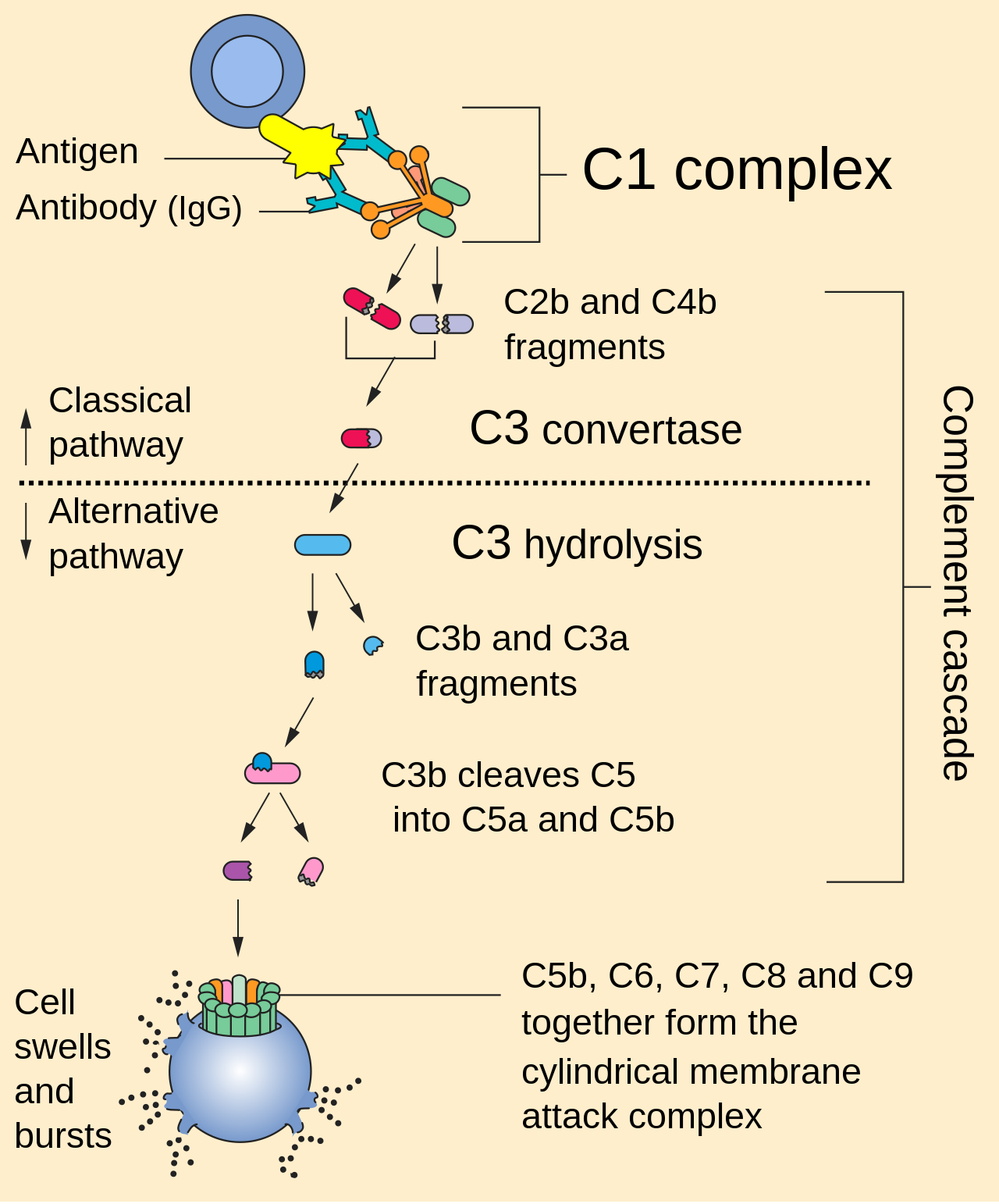
Complement Pathway
10.3.2 HIV as secondary immunodeficiency #
HIV is the worked case of acquired immunodeficiency because it removes the cell that organises adaptive immunity. gp120 binds CD4 and then a chemokine receptor, CCR5 on the memory CD4 cells and macrophages that transmit infection, or CXCR4 on strains that appear later in some patients. gp41 fuses the membranes. Macrophages and dendritic cells are infected too. Acute viraemia often causes a mononucleosis-like illness and a sharp fall in CD4 cells, partly reversed as CD8 cells respond. A set point of continuous replication in lymphoid tissue follows, and the CD4 count declines over years. The quiet clinical middle is not virological latency. Cells are lost by direct killing, by CD8 attack, and by death of bystander cells. AIDS, by the conventional definition, is a CD4 count below 200 cells per microlitre or a specified opportunistic disease. A healthy count is usually several hundred to well over a thousand, and the CD4:CD8 ratio of about two to one inverts. Opportunistic infection follows because help is gone: macrophages are not activated, germinal centres fail, CD8 memory is poorly kept, and mucosal Th17 cells are lost early. Pneumocystis pneumonia is the classic picture. Microbiology owns the organisms.
10.4 Key terms, summary, and self-check #
Key terms. Anaphylaxis; haemolytic disease of the newborn; serum sickness; tuberculin test; type 1 diabetes; Graves disease; SLE; SCID; common γ chain; X-linked agammaglobulinaemia; hyper-IgM; chronic granulomatous disease; CCR5; AIDS.
Summary. Type I is IgE and mast cells, type II is antibody on a surface, type III is immune complexes, and type IV is delayed T-cell inflammation. T cells destroy islet β cells in type 1 diabetes; stimulating antibody drives Graves disease; nucleic-acid antibodies and interferon drive lupus. SCID removes T and B function, Bruton tyrosine kinase removes B cells, the oxidase defect defines chronic granulomatous disease, and terminal complement deficiency removes lysis of Neisseria. HIV enters through CD4 and a chemokine receptor and, by deleting CD4 cells, removes the help that had contained organisms already met.
Self-check.
1. For each of types I–IV, name the effector and one example.
2. Why does haemolytic disease of the newborn destroy foetal red cells and not the mother’s?
3. What is destroyed in type 1 diabetes, and by which arm? How does the effector in Graves disease differ?
4. Match X-linked SCID, RAG deficiency, X-linked agammaglobulinaemia, chronic granulomatous disease, and terminal complement deficiency to the missing function.
5. Which cell does HIV deplete, which molecules does it use to enter, and why do opportunistic infections include organisms already met?
