
 SVG
SVGAntigen Presentation
Antigen presentation stimulates T cells to activate "cytotoxic" CD8+ cells or "helper" CD4+ cells. Cytotoxic cells directly attack other cells carrying certain foreign or abnormal molecules on their surfaces. Helper T cells, or Th cells, coordinate immune responses by communicating with other cells. In most cases, T cells only recognize an antigen if it is carried on the surface of a cell by one o
 SVG
SVGClonal Selection
Abstract diagram of the clonal selection of B and T lymphocytes. Legend: Hematopoietic stem cell Immature lymphocytes with various receptors "Self"-antigens from the body's own tissues Mature, inactive lymphocytes Foreign antigen Cloned activated lymphocytes
 SVG
SVGComplement Alternative
The molecular machinery of the central dogma governs genetic storage, replication, transcription, and translation in living systems.
 SVG
SVGComplement Leclassic
Neuronal electrical signaling relies on rapid, transient voltage reversals known as action potentials propagating along excitable axolemmas.
 SVG
SVGShowing The Structure Of An Immunoglobulin
The vertebrate immune system coordinates innate barrier defenses with antigen-specific adaptive immunity.
 SVG
SVGImmunoglobulin Basic Unit
Schematic diagram of the basic unit of immunoglobulin (antibody) Fab Fc heavy chain (consist of VH, CH1, hinge, CH2 and CH3 regions: from N-term) light chain (consist of VL and CL regions: from N-term) antigen binding site hinge regions (*) -S-S- mean disulfide bonds.
 SVG
SVGMhc Class 1
Schematic representation of MHC class I molecule, consisting of three α-domains and one β2-microglobulin molecule. The peptide-binding groove is situated between domains α1 and α2.
 SVG
SVGMhc Class 2
Schematic representation of MHC class II molecule, consisting of two α-domains and two β-domains. The peptide-binding groove is situated between domains α1 and β1.
 SVG
SVGIl-18 Signaling Pathway (Homo Sapiens)
Interleukin-18 (IL-18) is a member of the IL-1 family of cytokines and was initially described as an IFN-γ-inducing factor derived from anti-CD3-stimulated T-helper (Th)1 cells. IL-18 plays a significant role in the activation of hematopoietic cell types mediating both Th1 and Th2 responses and is the primary inducer of interferon-γ in these cells. The biological activity of IL-18 is mediated through its binding to the IL-18 receptor complex and activation of nuclear factor-κB (NF-κB), culminating in the production and release of several cytokines, chemokines, and cellular adhesion molecules. In certain cell types, IL-18 also activates mitogen-activated protein kinases (MAPKs) and phosphoinositide 3-kinase/ AKT serine/threonine kinase (PI3K/AKT) signaling modules leading to the production and release of proinflammatory cytokines. IL-18-mediated signaling acts as one of the vital components of the immunomodulatory cytokine networks involved in host defense, inflammation, and tissue regeneration. Albeit its biomedical importance, a comprehensive resource of IL-18 mediated signaling pathway is currently lacking. In this study, we report on the development of an integrated pathway map of IL-18/IL-18R signaling. The pathway map was developed through literature mining from published literature based on manual curation guidelines adapted from NetPath and includes information on 16 protein-protein interaction events, 38 enzyme-catalysis events, 12 protein translocation events, 26 activations/inhibition events, transcriptional regulators, 230 gene regulation events and 84 induced protein expression events.
 SVG
SVGTcr Complex
TCR complex A diagram showing the alpha-beta T cell receptor complex, including associated CD3 proteins. The blue segments are the ITAMs.
 SVG
SVGBcr (Nih Bioart 51 - 627919)
B cell receptor with colored binding sites. The B-cell receptor complex is made up of cell-surface immuno-globulin
 SVG
SVGBcr (Nih Bioart 51 - 627924)
B cell receptor with colored binding sites. The B-cell receptor complex is made up of cell-surface immuno-globulin
 SVG
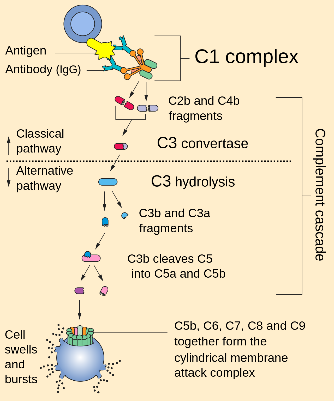
SVGComplement Pathway
The complement system is made up of about 25 proteins that work together to “complement” the action of antibodies in destroying bacteria. Complement proteins circulate in the blood in an inactive form. When the first protein in the complement series is activated— typically by antibody that has locked onto an antigen—it sets in motion a domino effect. Each component takes its turn in a precise chain of steps known as the complement cascade. The end product is a cylinder inserted into—and puncturing a hole in—the cell’s wall. With fluids and molecules flowing in and out, the cell swells and bursts.
 SVG
SVGImmune Checkpoints Of Immunosuppressive Actions Associated With Breast Cancer
Immune checkpoints of inhibitory pathways are fundamental in the immune system to maintain self-tolerance and modulate immune responses. In breast cancer, some of these immune checkpoints and immunosuppressive factors have been associated with subtype specificity through their expressions on breast cancer cells. The different immune cells and their ligand-receptor interactions and secreted stimulatory growth factors, chemokines and cytokines are shown. The expression of these immune markers in the TME in breast cancer may or not be subtype-specific but are important in circumventing immune recognition or to immobilize effector T cells. Thus, the expression of these ligands and receptors may be associated with breast cancer stage and clinical outcome. (AKT. serine/threonine kinase or protein kinase B; ARG1. Arginase 1; Bcl-xL. B-cell lymphoma-xtra large; CAFs. Cancer-associated fibroblasts; CTLA4. cytotoxic T-lymphocyte-associated protein 4; ER− Estrogen Receptor Negative; GM-CSF. Granulocyte-Macrophage Colony-Stimulating Factor; HER2+. Human Epidermal Growth Factor Receptor 2; IDO. Indoleamine-2,3-dioxygenase; IFN-γ. interferon gamma; IFN-γR. interferon gamma receptor; IL. Interleukin; JAK. Janus kinase; iNOS. Inducible nitric oxide synthase; MDSC. Myeloid-derived suppressor cells; MHC. major histocompatibility complex; MMP. Matrix Metalloproteinases; mTOR. Mammalian target of rapamycin; MUC. Mucins; NK. natural killer; NF-κB. nuclear factor-κB; NLR. Neutrophil-lymphocyte ratio; NO. Nitric Oxide; PI3K. PI3K.phosphoinositide 3-kinase; PD-1. programmed death-1; PD-L1. Programmed death-ligand1; PGE2. Prostaglandin E2; PR−. Progesterone Receptor Negative; ROS. Reactive Oxygen Species; SHP. Src homology protein-tyrosine phosphatase; Siglec 9. Sialic acid-binding lectins 9; STAT. Signal transducer and activator of transcription; TAMs. Tumor-associated macrophages; TANs. Tumor-associated neutrophils; TGF-β. Transforming Growth Factor-beta; TIL. Tumor-Infiltrating Lymphocytes; TNBC. Triple Negative Breast Cancer; Treg. regulatory T cell; TCR. T cell receptor; VEGF. Vascular endothelial growth factor). [ 1 ]
 SVG
SVGImmune Response Of Lymphocytes
APCs present antigen on their Class II MHC molecules Helper T cells recognize these and macrophages continue the proliferation of T helper cells.
 SVG
SVGT Cell Activation Ku
Kurdishː Li ser rûyê hokarên nexweşiyê de dijepeydaker heye. Li ser rûyê lîmfexaneyên T jî proteînên wergir hene. Heke lîmfexane rastê dijepeydaker were, bi alikariya proteînên wergir dijapeydakerê dinase û xwe bi dijepeydakerê ve girê dide. Anko cureyek xaneya T, yek cureya proteînên wergir ên diyarkirî li ser parzûna xaneya xwe digirin. Gava lîmfexane cara pêşîn rastê hokara nexweşiyê tê, wê gavê li dij hokara nexweşiyê çalak dibe. Xaneya T her tim raste rast bi hokara nexweşiyê ve tekilî çê neke, di nav laş de bi navê “xaneyên desnîşankirina dijepeydaker” hin xane hene(antigen presenting cell), van xaneyan alîkariya xaneyen T dikin ku ji hebûna hokara nexweşiya ji derveyê laş hatiye agahdar be. Li ser rûyê xaneyên desnîşankirina dijepeydaker de hin proteînên taybet hene ev proteîn wekî pêkhateya MHC (major histocompatibility complexes) tê binavkirin. Dijepeydaker xwe li ser MHC girê dide, xaneyên T jî bi vî awayê ji wan agahdar dibe. MHC du cure ne, MHC class I û MHC class II. Gellek cureyên xaneyên desnîşankirina dijepeydaker heye, xaneya hellûşênera gewre yek ji van xaneyan e. Xaneya hellûşênera gewre (xaneya makrofaj) beşek ji bergiriya giştî ye, lê ji boy bergiriya taybet jî kar dike. Hellûşênera gewre hokara nexweşiyê qût dide (diqurtîne) û di nav xaneyê de diherisîne. Piştê hilweşîna hokara nexweşiyê, hin pirtikên(perçeyên) dijepeydaker li ser rûyê parzûna xaneya hellûşênera gewre de, li ser proteînên MHC de cih dibin. Anko xaneya desnîşankirina dijepeydaker, hokara nexweşiyê(dijepeydaker) li ser protnên MHCyan de bi xaneyên T ve dide naskirin. Ev pirtikên li ser rûyê hellûşênera gewre xaneyên T çalak dikin. Xaneyên T xwe bi pirtikên dijepeydakerên li ser MHCyan ve girê didin. Dibe ku ji xeynî xaneyên hellûşênera gewre, pirtikên dijepeydaker li ser rûyê xaneyên tûşî hokara nexweşiyê bûyî jî hebin. Vê demê xaneyên T xwe bi van xaneyên tûşbûyî ve jî girê didin.
 SVG
SVGPhagocytosis Zp
This detailed scientific diagram illustrates Phagocytosis Zp in the context of Immunology.