
4.1 Semiconservative copying #
4.1.1 Meselson and Stahl #
Conservative replication would preserve the parental duplex and build an entirely new one. Semiconservative replication gives each daughter one parental strand and one new strand. Dispersive replication would scatter parental material through both daughters.
Meselson and Stahl (1958) grew E. coli for many generations in 15N, then switched the cells to 14N and followed DNA density in a caesium-chloride gradient.
Conservative replication is ruled out by the first generation: it would have left one heavy band and one light band. Dispersive replication is ruled out by the second: it would have produced one band, drifting lighter, not two discrete bands.
4.2 Where a fork starts #
The study of Where a fork starts provides fundamental principles governing cellular architecture, molecular interactions, and physiological regulation.

Replication Fork
4.2.1 oriC and DnaA #
E. coli has one origin, oriC, about 245 bp, with DnaA-binding sites and an AT-rich unwinding element. Two forks leave in opposite directions and meet in the terminus region, where topo IV decatenates the daughters.
Initiation is held to once per cycle by the availability and ATP state of DnaA, and by sequestration of the newly replicated, hemimethylated origin. The principle is the same in eukaryotes.
4.2.2 ORC in eukaryotes #
Eukaryotic chromosomes are long and have many origins. In budding yeast the origins are defined sequence elements.
The origin recognition complex (ORC) binds potential origins. In G1, ORC with Cdc6 and Cdt1 loads the MCM helicase as an inactive double hexamer. That loading is licensing: the origin may fire later, and it is not yet firing. S-phase CDK and the DDK kinase Cdc7 activate MCM as the CMG helicase (Cdc45, MCM, GINS). Not every licensed origin fires every S phase. Early origins tend to lie in euchromatin and late origins in heterochromatin.
4.3 The replication fork #
The study of The replication fork provides fundamental principles governing cellular architecture, molecular interactions, and physiological regulation.

Dna Replication Numbered
4.3.1 Helicase, single-strand binding protein, and primase #
Helicase unwinds the duplex. Single-strand binding protein keeps the strands apart and protects them: SSB in bacteria, RPA in eukaryotes. Primase lays down a short RNA primer, because DNA polymerases cannot start a chain. They can only extend a 3′-OH.

Xbio Illustration Dna Helicase
Directions differ and are worth fixing once. Bacterial DnaB moves 5′→3′ on the lagging-strand template. Eukaryotic CMG moves 3′→5′ on the leading-strand template. Pol α does not proofread, so the accurate polymerases take over.
4.3.2 Why synthesis runs 5′→3′ #
The 3′-OH of the last nucleotide attacks the 5′ triphosphate of the incoming nucleotide, and pyrophosphate leaves. The chain grows 5′→3′ while the template is read 3′→5′. No cellular DNA polymerase grows a chain 3′→5′.
The restriction is what makes proofreading possible. A wrong nucleotide is removed from the 3′ end, and the end that remains is again a 3′-OH. A polymerase that added nucleotides at the 5′ end would carry the activating triphosphate on the growing chain. Cutting off a mistake would leave a 5′ monophosphate that cannot continue. Proofreading and chain growth would be chemically at odds. Both strands of the fork are therefore made 5′→3′, even though they point opposite ways.
4.3.3 Leading strand, lagging strand, and Okazaki fragments #
The fork moves in one direction. The leading strand is copied continuously, one primer at the origin and then uninterrupted synthesis in the direction of fork movement.
In bacteria the fragments are about 1,000–2,000 nucleotides. In eukaryotes they are about 100–200 nucleotides, near nucleosome length. The lagging strand loops, in the trombone arrangement, so that both polymerases can face the same way and still synthesise 5′→3′. Discontinuity is the geometry of antiparallel strands plus 5′→3′ chemistry. It is not a defect of the enzyme.
4.3.4 DNA polymerase III versus pol δ and pol ε #
Names collide here. DNA polymerase I of E. coli is not RNA polymerase I, and DNA polymerase III is not RNA polymerase III. Its core polymerises with the α subunit and proofreads with the ε subunit, the product of dnaQ. A mutation in ε is a mutator, which is genetic evidence that proofreading happens in the cell.

Pcna Dna Polymerase Dna
More than one core works at a fork. The classical picture is a dimer, one core on each strand. The leading-strand enzyme does not turn round.
Eukaryotes divide the labour three ways. Pol α primes and hands over the 3′ end.
4.3.5 PCNA and the sliding clamp #
A polymerase core falls off after a short stretch. A sliding clamp, a ring around the duplex, tethers it.
A clamp loader opens the ring and shuts it on a primer–template junction, using ATP. The bacterial loader is the γ complex. Pol δ, FEN1, DNA ligase I, and several repair proteins bind it. Loading has a direction, and that direction later helps mismatch repair tell which strand is new (Chapter 5).
4.3.6 Proofreading by the 3′→5′ exonuclease #
The polymerase active site already rejects most wrong nucleotides, to an error of about 10^−4 to 10^−5 per base. A mismatched 3′ end frays, moves to the exonuclease site, and is removed. Proofreading improves fidelity by about a hundredfold, toward about 10^−7. Mismatch repair later supplies another large factor, toward about 10^−10 overall.
The exonuclease runs 3′→5′, the opposite of synthesis, and it works only because synthesis itself is 5′→3′. Pol α has no such activity, which is one reason its primer must be replaced. That is a fact about the enzyme, not a method.
4.3.7 Removing the primer: RNase H, FEN1, and ligase #
Every fragment starts with RNA, and RNA does not belong in the finished chromosome.
In E. coli, DNA polymerase I removes the primer. Its 5′→3′ exonuclease nibbles RNA at a nick while its polymerase fills behind, a nick translation. It does not synthesise the bulk of the chromosome.
In eukaryotes, pol δ runs into the previous fragment and peels its 5′ end up as a flap. FEN1 cuts at the base of a short flap. RNase H degrades RNA in an RNA–DNA hybrid and can remove most of a primer, often leaving the last ribonucleotide for FEN1. Long flaps are trimmed with help from Dna2 before the final cut. DNA ligase I, brought by PCNA, seals the nick, using ATP rather than NAD+.
4.4 Ends of linear chromosomes #
4.4.1 The end-replication problem #
A circular bacterial chromosome has no end. The last primer is just another nick, filled from upstream DNA. On the lagging strand, removal of the terminal RNA primer leaves a gap, and there is no upstream 3′-OH to fill it. Conventional DNA polymerases cannot start at the tip.

Xbio Illustration Dna Replication Fork
The leading-strand end is related. Synthesis can run to the last nucleotide and leave a blunt end, but telomeres are not kept blunt. Nucleases resect the 5′ strand to restore a 3′ overhang, so both ends lose sequence if nothing restores it. Hayflick had already seen the cellular consequence without the molecule: normal human somatic cells in culture divide a finite number of times, classically on the order of fifty population doublings for fibroblasts, and then stop.
4.4.2 Telomeres and telomerase #
Telomeres are repetitive ends plus proteins. The human repeat is TTAGGG, for several kilobases, with a G-rich 3′ overhang. Shelterin, including TRF1, TRF2, and POT1, coats the end so the cell does not treat it as a double-strand break.

Telomere Scheme
Telomerase is a reverse transcriptase. The protein TERT copies an internal RNA, TERC, complementary to the telomeric repeat, and extends the 3′ overhang. The logic is the same and the sequence is not the human one. Telomerase does not copy the rest of the chromosome, and it does not replace proofreading.
Telomerase is active in the germ line, in many stem cells, and in most cancer cells, which is one reason those clones escape the Hayflick limit. It is low or absent in most differentiated somatic cells. It is not sufficient, by itself, for transformation. It is not a complete theory of ageing.
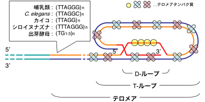
Telomere Structure
4.5 Key terms, summary, and self-check #
Key terms. Semiconservative; oriC; DnaA; ORC; MCM; Okazaki fragment; pol III; pol ε; pol δ; PCNA; FEN1; telomerase; Hayflick limit.
Summary. One intermediate-density band after one generation, and intermediate plus light DNA after two, establish semiconservative replication. DnaA opens oriC and DnaB is loaded; ORC and its loaders license eukaryotic origins by placing MCM once per cycle. All synthesis is 5′→3′ because only that polarity survives proofreading, so the lagging strand is made as Okazaki fragments. Bacterial pol III, and eukaryotic pol ε and pol δ, do the bulk copying; the β clamp or PCNA holds them on DNA. Primers are removed by pol I in bacteria and by RNase H, flaps, and FEN1 in eukaryotes, and ligase seals the nick. Linear ends cannot be completed by that chemistry. Telomerase extends the 3′ overhang from an internal RNA template, and shelterin hides the end.
Self-check.
1. Which Meselson–Stahl result eliminates conservative replication, and which eliminates dispersive replication?
2. What does DnaA do at oriC, and what is loaded at a eukaryotic origin in G1 that must not be loaded again once S phase has begun?
3. Why is the lagging strand discontinuous, and why could a polymerase that added nucleotides at the 5′ end not proofread and then continue?
4. Which eukaryotic polymerase primes, which copies most of the leading strand, and which ring makes pol δ processive?
5. What gap remains when the terminal RNA primer is removed, and does telomerase copy the chromosome or its own RNA?
