
5.1 Cell walls #
The study of Cell walls provides fundamental principles governing cellular architecture, molecular interactions, and physiological regulation.

Nucleus Of A Cell ()
5.1.1 Why a wall #
A wall is a secreted extracellular layer that resists turgor and gives shape. It is not a membrane: it is porous to small molecules, and the selective barrier remains the plasma membrane underneath. Cells that live in dilute water (bacteria, algae, plants, fungi) almost always have one. Animal cells do not. They use an extracellular matrix that is structural but not a rigid box, and they regulate volume with ion pumps instead of with a wall.

Plant Cell Wall
5.1.2 The plant wall #
The primary wall, laid down while the cell is still growing, is a composite.

Plant And Animal Cell Cytokinesis
- Cellulose microfibrils, bundles of β-1,4-glucan chains, are the tensile elements. They are spun by cellulose-synthase complexes in the plasma membrane, which move as they extrude, often guided by underlying cortical microtubules. The orientation of the newest microfibrils helps decide the direction in which the cell will expand.
- Hemicelluloses (for example xyloglucan) cross-link microfibrils.
- Pectins are acidic polysaccharides that form a hydrated gel and are abundant in the middle lamella, the glue between neighbouring cells.
- Proteins, including expansins, loosen the network so that turgor can stretch the wall during growth.
The secondary wall is deposited inside the primary wall after growth stops, in cells such as xylem vessels and fibres. It is thick, cellulose-rich, and often impregnated with lignin, a phenolic polymer that makes wood rigid and waterproof. Plasmodesmata, the cytoplasmic channels, are kept open through both walls.
A plant protoplast, the cell with its wall digested off by enzymes, is spherical and osmotically fragile. That experiment assigns turgor resistance to the wall.
5.1.3 Fungal and bacterial walls, for comparison #
Fungal walls use chitin (β-1,4-linked N-acetylglucosamine) and β-glucans. They do not contain cellulose in the plant pattern, and they do not contain peptidoglycan. The echinocandin antifungal drugs inhibit glucan synthesis; they have no target in human cells, which is the pharmacological point of a wall humans do not possess.
Bacterial peptidoglycan was described in Chapter 1. The teaching comparison is chemical: plants use cellulose, fungi use chitin, bacteria use peptidoglycan, and each is a target that the others lack.
5.2 The animal extracellular matrix #
The study of The animal extracellular matrix provides fundamental principles governing cellular architecture, molecular interactions, and physiological regulation.

Extracellular Matrix
5.2.1 Fibres, gel, and adhesive glycoproteins #
The extracellular matrix (ECM) is everything a tissue secretes into the space outside the plasma membrane. Its composition differs by tissue. Cartilage, tendon, bone, and the thin sheet under an epithelium are all ECM, built from different proportions of the same families.

N Linked Protein Glycosylation In The Er
- Collagen is the main tensile fibre. The primary sequence is a repeating Gly-X-Y, often with proline and hydroxyproline in X and Y. Three chains wind into a triple helix, and those molecules pack into fibrils (in the fibrillar collagens, types I, II, and III). Vitamin C is required for the hydroxylases. Scurvy, a failure of collagen, opens old wounds and loosens teeth because new collagen cannot be stabilised. Type IV collagen does not form fibrils; it forms the network of the basal lamina.
- Elastin, cross-linked into fibres with fibrillin, recoils after stretch. It is abundant in arteries, lungs, and elastic ligaments.
- Proteoglycans are core proteins carrying glycosaminoglycan (GAG) chains, which are long, negatively charged, and bind water. They make the gel that resists compression. Aggrecan in cartilage is the extreme case. Hyaluronan is a GAG that is not attached to a core protein and can be enormous.
- Adhesive glycoproteins link cells to the rest. Fibronectin has an RGD sequence (arginine-glycine-aspartate) that integrins bind. Laminin is the organiser of the basal lamina and binds integrins, collagen IV, and other matrix molecules.
5.2.2 Basal lamina #
The basal lamina is a thin, specialised ECM under epithelia and around muscle, fat, and Schwann cells. Laminin, type IV collagen, nidogen, and the heparan-sulfate proteoglycan perlecan are its core. It separates an epithelium from the underlying connective tissue, filters molecules (the kidney glomerulus), and tells the attached cells where they are. Cells that lose contact with it, or that are told by mutation that contact is unnecessary, behave as if they were free to migrate. That fact returns in the discussion of transformed cells.
5.3 Junctions #
5.3.1 Cell–cell junctions of animal tissues #
Epithelia show the full set, arranged from the apical side downward as a junctional complex.
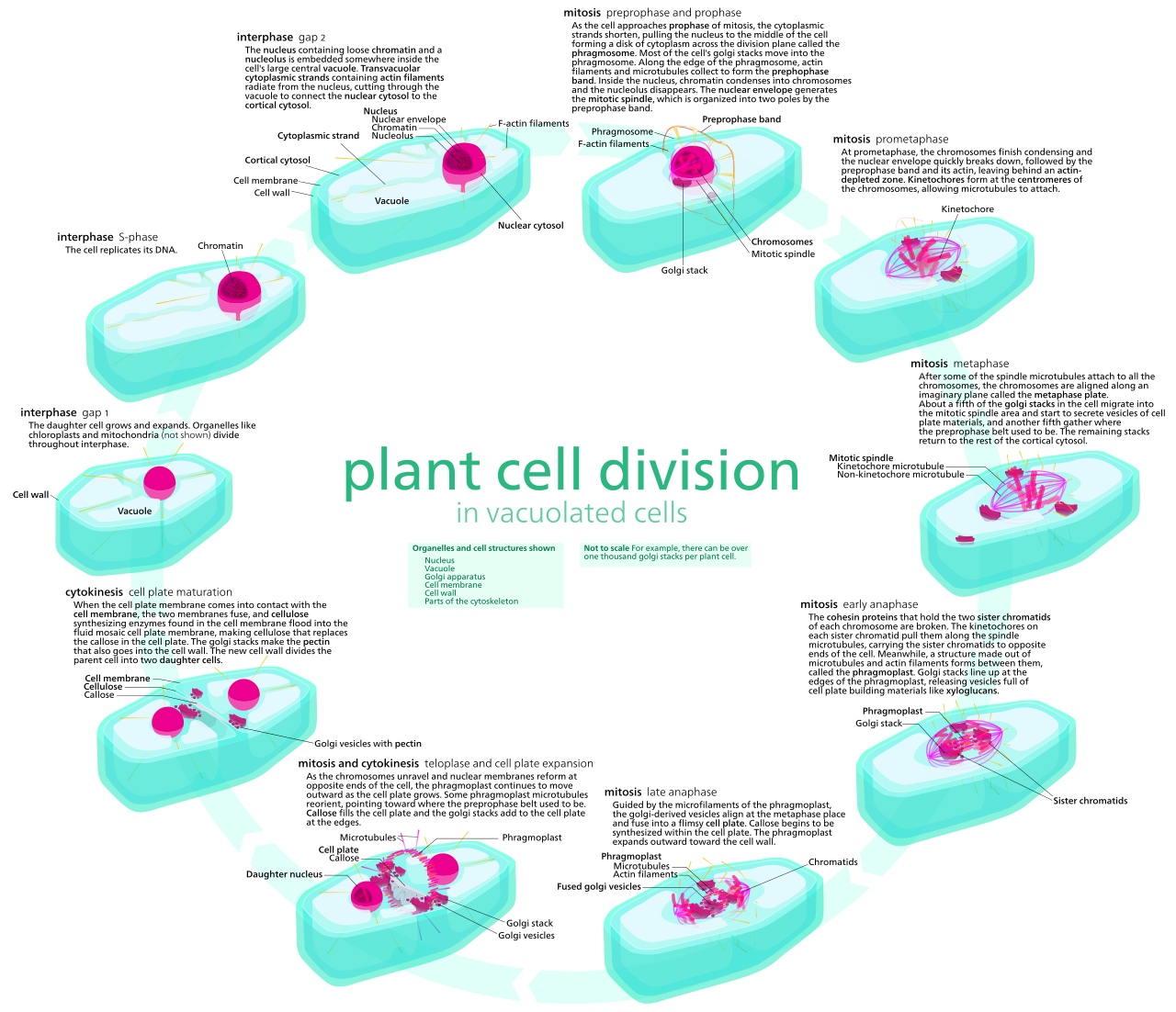
Plant Cell Cycle
| Junction | Principal adhesion proteins | Cytoskeleton attached | Job |
|---|---|---|---|
| Tight junction (zonula occludens) | Claudins, occludins; ZO proteins inside | Actin | Seals the space between cells so that solutes must cross the epithelium through cells; also fences membrane proteins so that apical and basolateral proteins do not mix |
| Adherens junction | Classical cadherins; catenins inside | Actin | Mechanical adhesion; the belt of adherens junctions lets a sheet of cells change shape together, as in forming a tube |
| Desmosome | Desmoglein and desmocollin (desmosomal cadherins); plakoglobin, desmoplakin | Intermediate filaments | Spot welds that resist shear. Abundant in skin and cardiac muscle |
| Gap junction | Connexins; six make a connexon, two connexons make the channel | None as a mechanical link | A pore that passes ions and metabolites up to about 1,000 daltons. Electrically and metabolically couples the cells. Closes when Ca2+ rises or pH falls, isolating a damaged neighbour |
Cadherins need extracellular Ca2+. Remove calcium and adherens junctions and desmosomes let go, which is why tissues are dissociated for culture in calcium-free conditions with a protease. Cadherins bind the same cadherin on the neighbouring cell (homophilic binding). This is one molecular basis of cells sorting into like tissues.
Gap junctions in invertebrates use innexins, not connexins. The function is the same. Plants do not use gap junctions; plasmodesmata carry that role and can pass much larger molecules, including some proteins and RNAs, because the channel is a plasma-membrane-lined pore with an ER tubule (the desmotubule) inside it.
5.3.2 Cell–matrix junctions #
Focal adhesions attach actin stress fibres to the ECM through integrins. Integrins are heterodimers of an α and a β subunit. Binding ligand on the outside changes the conformation and allows talin, vinculin, and other proteins to connect the cytosolic tail to actin. Force strengthens the attachment. Integrins also signal: “I am attached” is information the cell uses when deciding to divide or to die. Many cells undergo apoptosis if they are held in suspension (anoikis). Attachment is a survival signal.

Extracellular Matrix V1.001 Labeled
Hemidesmosomes attach intermediate filaments to the basal lamina through integrin α6β4 and are the stable anchors of an epithelium. Mutations in the relevant keratins, laminin, or integrin cause blistering diseases, because the epithelium tears off its base under ordinary friction.
5.3.3 Polarity as a consequence of junctions #
Once tight junctions fence the membrane and the cytoskeleton is anchored, a cell can keep different transporters on its apical and basolateral faces. The intestinal glucose system of Chapter 3 depends on this. Junctions are not only glue. They are how a tissue becomes a directional machine.

Mitosis Cells Sequence
5.4 Key terms, summary, and self-check #
Key terms. Cellulose; pectin; middle lamella; lignin; plasmodesmata; collagen; proteoglycan; RGD; integrin; cadherin; claudin; connexin; basal lamina; focal adhesion; hemidesmosome.
Summary. Walls resist turgor with different polysaccharides in plants, fungi, and bacteria. Animal tissues use a composite ECM of collagen, elastin, proteoglycans, and adhesive glycoproteins. Tight junctions seal and fence, adherens junctions and desmosomes bear mechanical load through actin and intermediate filaments, gap junctions couple cytoplasms, and integrins connect the ECM to the cytoskeleton and to survival signalling.
Self-check.
1. Why does digesting the plant cell wall produce a spherical, fragile protoplast?
2. A pathologist sees epithelium lifting off the basal lamina in a blister. Which class of junction has failed, and which cytoskeletal polymer was it connected to?
3. How can a small metabolite pass from one epithelial cell to the next without crossing the extracellular space, and what size limit applies?
4. Why is vitamin C deficiency a collagen disease?
5. Claudin loss makes an epithelium leak. Does it also let apical transporters drift to the basolateral membrane? Explain.
