
4.1 The nucleus #
The study of The nucleus provides fundamental principles governing cellular architecture, molecular interactions, and physiological regulation.

Nucleus Er Golgi
4.1.1 Envelope, lamina, and pores #
The nuclear envelope is a double membrane. The outer membrane is continuous with the rough ER and often carries ribosomes. The space between the membranes is continuous with the ER lumen. The inner membrane is lined by the nuclear lamina, a mesh of lamin intermediate filaments that supports the envelope and binds chromatin. Mutations in lamin A cause Hutchinson–Gilford progeria, a premature-aging disease, which shows that the lamina is structural and regulatory, not just a scaffold.
Traffic in and out goes through nuclear pore complexes. A vertebrate pore is a huge assembly, on the order of 100–125 MDa, built from nucleoporins. Molecules smaller than roughly 40 kDa can diffuse through slowly. Larger proteins, and RNAs packaged as particles, are carried by receptors. The rules of that transport are in Chapter 9. During open mitosis in animal cells the envelope breaks into vesicles and the lamina is disassembled by phosphorylation, then both reassemble around the daughter chromosomes.
4.1.2 Chromatin and the nucleolus #
DNA in the nucleus is packaged with histones into nucleosomes (“beads on a string”) and folded further into higher-order chromatin. Euchromatin is relatively open and is where most transcription occurs. Heterochromatin is compact, often at the nuclear periphery and around centromeres and telomeres, and is largely silent. Chromosomes occupy territories rather than being stirred at random. The packing is dynamic: a gene that switches on often moves to a more open region.
The nucleolus is the factory for ribosomal subunits. It forms around nucleolar organiser regions, the chromosomal stretches that carry tandem repeats of ribosomal RNA genes. Ribosomal RNA is transcribed and processed there, and ribosomal proteins imported from the cytosol assemble with it. The subunits leave through nuclear pores and become active only in the cytosol, which prevents half-made ribosomes from translating nuclear RNA. A cell that is making protein rapidly has a prominent nucleolus. That is a visible readout of growth.
4.2 Endoplasmic reticulum #
4.2.1 Rough ER #
Rough ER is a network of flattened sacs studded with ribosomes on the cytosolic face. Those ribosomes are not a special population. A ribosome binds the ER only while it is making a protein that carries an ER signal sequence (Chapter 7). The rough ER is the entry point of the secretory pathway: secreted proteins, plasma-membrane proteins, and the proteins of the ER, Golgi, lysosomes, and endosomes all start here, or are inserted into this membrane.
4.2.2 Smooth ER #
Smooth ER is tubular and has no ribosomes. Its jobs depend on the cell.
- Lipid synthesis. Membrane phospholipids, cholesterol, and ceramide are made by enzymes facing the cytosol. Cells that secrete steroid hormones, such as those of the adrenal cortex and the gonads, have extensive smooth ER because the pathway is membrane-bound.
- Detoxification. Hepatocytes expand the smooth ER when challenged with drugs such as phenobarbital. Cytochrome P450 enzymes oxidise hydrophobic compounds, making them more soluble and easier to excrete. The same enzymes activate some compounds into toxins.
- Calcium storage. The sarcoplasmic reticulum of muscle is a specialised smooth ER. The SERCA pump loads it with Ca2+, and a nerve impulse releases Ca2+ through ryanodine receptors to trigger contraction. Non-muscle cells use the ER the same way, on a smaller scale, through IP3 receptors.
The rough and smooth ER are continuous. A protein or a lipid made in one region can diffuse to the other.
4.3 Golgi apparatus #
The Golgi apparatus is a stack of flattened cisternae, polarised from cis (entry) to trans (exit), with a trans-Golgi network as the sorting station. In animal cells the stack usually sits near the centrosome. In plant cells many separate stacks are distributed through the cytoplasm.

Disrupted Golgi (Nih Bioart 627)
Material arrives from the ER at the cis face, travels through the stack, and leaves from the trans-Golgi network toward the plasma membrane, the endosomes and lysosomes, or secretory granules. Two ideas describe the travel. In the vesicular-transport model, cisternae are stable and cargo moves forward in vesicles. In the cisternal-maturation model, a cisterna itself moves forward and matures, while resident enzymes recycle backward. Current evidence favours maturation as the main path for large cargo, with vesicles doing the recycling. Both vesicle coats appear in Chapter 8.
What the Golgi adds, chemically:
- processing of N-linked oligosaccharides from the high-mannose form received from the ER toward complex forms, with cell-type-specific additions of galactose, fucose, and sialic acid;
- O-linked glycosylation of selected serines and threonines;
- assembly of the glycosaminoglycan chains of proteoglycans;
- synthesis of sphingomyelin and glycolipids;
- sorting, which is the decision that makes a lysosomal enzyme, a secretory hormone, and a plasma-membrane protein leave in different vesicles.

Golgi Apparatus (Numbers Version)
4.4 Lysosomes #
The study of Lysosomes provides fundamental principles governing cellular architecture, molecular interactions, and physiological regulation.

Lysosomes Digestion
4.4.1 Acid hydrolases behind a sealed membrane #
A lysosome is a single-membrane organelle packed with hydrolytic enzymes (proteases, nucleases, lipases, glycosidases) that work best near pH 4.5–5. A V-type proton pump in the membrane maintains that pH. The membrane itself is protected by heavy glycosylation of its luminal proteins, which resists the hydrolases. If the enzymes leaked into the cytosol at pH 7 they would be far less active, which is one reason a ruptured lysosome does not automatically digest the cell. Massive rupture does injure the cell, and released cathepsins can participate in cell death.
Lysosomes receive cargo from endocytosis, phagocytosis, and autophagy. A primary lysosome has not yet met its substrate. After fusion it is a secondary lysosome. Undigested remnants can remain as residual bodies, prominent in long-lived cells.
4.4.2 Failure of lysosomal digestion #
Lysosomal storage diseases arise when one hydrolase is missing or mistargeted, so its substrate accumulates.

Apoptosis Pathways In Lysosomal Stress Response
- Tay–Sachs disease. Hexosaminidase A is deficient. The ganglioside GM2 accumulates in neurons, which is destructive because neurons are long-lived and rich in this lipid.
- I-cell disease. The enzyme that tags hydrolases with mannose 6-phosphate is deficient. The hydrolases are secreted instead of being sorted to lysosomes, and undegraded material builds up in inclusions (“inclusion cells”). This disease is the genetic proof of the sorting signal described in Chapter 8.
4.5 Peroxisomes #
Peroxisomes are single-membrane organelles, often with a crystalline core of oxidase. They are named for hydrogen peroxide, which they both make and destroy.
Oxidases remove hydrogen from organic substrates and hand it to O₂, producing H₂O₂. Catalase then decomposes H₂O₂ to water and oxygen, or uses it to oxidise other substrates. Containing this chemistry in an organelle keeps a reactive oxidant out of the cytosol.
Other conserved jobs:
- β-oxidation of very-long-chain fatty acids, shortening them so that mitochondria can finish the job. In plant seeds, specialised peroxisomes called glyoxysomes run the glyoxylate cycle and convert stored fat to carbohydrate.
- Synthesis of plasmalogens, ether phospholipids that are abundant in myelin.
- Part of photorespiration in leaves. Glycolate produced by the oxygenase activity of Rubisco is metabolised in peroxisomes and mitochondria.
Peroxisomes import folded proteins from the cytosol and can form de novo as well as by growth and division. The import signals are in Chapter 9. Defects in the import machinery cause Zellweger syndrome, in which peroxisomes fail to assemble and very-long-chain fatty acids and plasmalogen metabolism are both disrupted.
4.6 Mitochondria #
The study of Mitochondria provides fundamental principles governing cellular architecture, molecular interactions, and physiological regulation.

Mercs Mitochondria Er Contact Sites ()
4.6.1 Four compartments #
A mitochondrion has an outer membrane, an inner membrane, the intermembrane space, and the matrix.
- The outer membrane contains porins (VDAC) and is permeable to ions and metabolites below about 5 kDa. It is not the permeability barrier that matters for energy conversion.
- The inner membrane is folded into cristae, rich in the phospholipid cardiolipin, and impermeable to protons and to most ions. It holds the respiratory chain and ATP synthase. The area of cristae scales with the respiratory demand of the tissue: heart muscle has dense cristae; a less active cell has fewer.
- The intermembrane space is chemically close to the cytosol for small molecules, because of the porins, but it holds proteins of its own, including cytochrome c.
- The matrix contains the enzymes of the citric-acid cycle (except succinate dehydrogenase, which is in the inner membrane), mitochondrial DNA, and mitochondrial ribosomes.
Mitochondria fuse and divide, and they move along microtubules. A textbook oval is a snapshot of a network.
4.6.2 From pyruvate to a proton gradient #
Glycolysis in the cytosol converts glucose to two molecules of pyruvate and yields a net of 2 ATP and 2 NADH per glucose. Pyruvate enters the matrix through a carrier. Pyruvate dehydrogenase decarboxylates it to acetyl-CoA. The citric-acid cycle oxidises acetyl-CoA to two CO₂ and reduces 3 NAD+ and 1 FAD per turn, also producing 1 GTP (or ATP) by substrate-level phosphorylation.
NADH and FADH₂ are reoxidised by the electron-transport chain in the inner membrane.
| Complex | What it does | Pumps protons? |
|---|---|---|
| I, NADH dehydrogenase | Takes electrons from NADH, passes them toward ubiquinone | Yes |
| II, succinate dehydrogenase | Takes electrons from succinate (FADH₂) and feeds ubiquinone; this is also a citric-acid-cycle enzyme | No |
| III, cytochrome bc1 | Transfers electrons from ubiquinol to cytochrome c | Yes |
| IV, cytochrome oxidase | Transfers electrons to O₂, forming water | Yes |
| ATP synthase (sometimes called complex V) | Lets protons back into the matrix and uses the flow to make ATP | Uses the gradient; does not pump in the forward direction |
Peter Mitchell’s chemiosmotic theory (1961) is the organising idea. Electron transport pumps protons out of the matrix. The inner membrane does not let them leak back. The resulting proton-motive force is the sum of the membrane potential (inside negative) and the pH gradient (inside alkaline). ATP synthase lets protons return through a rotor (the F₀ portion) and the rotation drives ATP synthesis on the F₁ head in the matrix. Uncouplers such as DNP, or the regulated uncoupling protein of brown fat, let protons back without ATP synthesis. Electron transport then runs, fuel is burned, and the energy leaves as heat.
A modern estimate of the yield is about 2.5 ATP per mitochondrial NADH and about 1.5 ATP per FADH₂, not the older integers 3 and 2. The yield from cytosolic NADH depends on the shuttle that moves the reducing power in: the malate–aspartate shuttle preserves it as NADH, and the glycerol-phosphate shuttle hands it to ubiquinone at the FADH₂ level. A realistic net for complete oxidation of glucose in a eukaryote is about 30 ATP, varying with shuttle and with proton leak. Quoting 36 or 38 repeats an outdated stoichiometry.
4.6.3 What else mitochondria do #
Mitochondria are not only ATP factories.
- They buffer and release Ca2+, in conversation with the ER.
- They make iron–sulfur clusters and parts of the haem pathway.
- They decide part of apoptosis. Proteins of the Bcl-2 family control pores in the outer membrane; cytochrome c released into the cytosol helps assemble the apoptosome (Chapter 13).
- Their own genome means mitochondrial diseases exist, often affecting muscle and nerve, the tissues with the highest ATP demand, and inheritance is maternal in the usual case.
A cell without mitochondrial DNA can survive in culture if the medium replaces what respiration would have supplied: a fermentable sugar, and usually pyruvate and uridine. It cannot respire. Those rho-zero cells are an experimental tool.
4.7 Chloroplasts #
The study of Chloroplasts provides fundamental principles governing cellular architecture, molecular interactions, and physiological regulation.
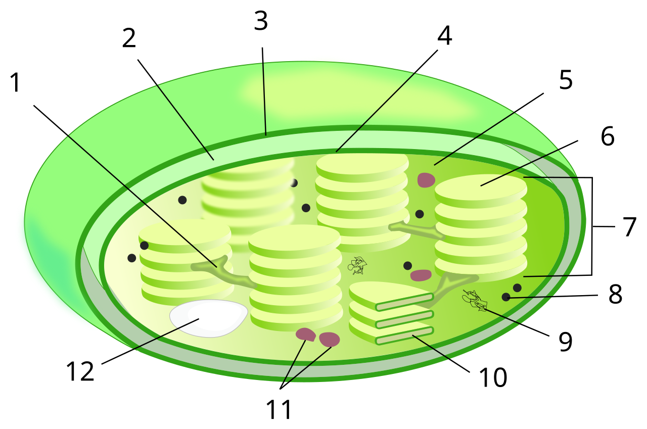
Chloroplast Numb
4.7.1 Compartments and plastid family #
Chloroplasts have a double envelope and a third membrane system, the thylakoids. Stacks of thylakoids are grana, joined by stroma lamellae. The interior of the thylakoid is the thylakoid lumen. The fluid outside the thylakoids, inside the inner envelope, is the stroma.
Chloroplasts are one differentiation state of plastids. Proplastids in dividing cells become chloroplasts in the light, amyloplasts where starch is stored, or chromoplasts in ripening fruit and flowers. They can convert from one form to another. All of them in a given plant share the same plastid genome, typically 120–160 kilobases and on the order of 100 genes in flowering plants.
4.7.2 Light reactions and the Calvin cycle, as cell biology #
The light reactions are a chemiosmotic machine built on the same principle as the respiratory chain, with the polarity reversed in one important sense: protons are pumped into the thylakoid lumen, and ATP synthase lets them out into the stroma, where the ATP is needed.

Life Cycle And Protein Associations Of Connexins
The route of electrons in the linear (non-cyclic) pathway, the Z-scheme:
1. Photosystem II, with reaction centre P680, absorbs light and pulls electrons from water. The oxygen-evolving complex releases O₂ and protons into the lumen.
2. Electrons travel through plastoquinone, the cytochrome b₆f complex (which pumps protons), and plastocyanin.
3. Photosystem I, reaction centre P700, absorbs a second photon and lifts the electrons to a more negative potential.
4. Ferredoxin and ferredoxin–NADP+ reductase reduce NADP+ to NADPH in the stroma.
Cyclic electron flow around photosystem I makes extra ATP without NADPH or O₂, which cells use when the Calvin cycle demands more ATP than the linear pathway supplies.
The Calvin cycle in the stroma fixes CO₂. Rubisco adds CO₂ to ribulose 1,5-bisphosphate, producing two molecules of 3-phosphoglycerate. ATP and NADPH from the light reactions reduce this to triose phosphate, and the cycle regenerates the acceptor. Rubisco also reacts with O₂ (photorespiration), which wastes carbon. C₄ and CAM plants concentrate CO₂ at Rubisco by prior fixation into a four-carbon acid, a structural solution to a kinetic problem of one enzyme. The leaf peroxisomes and mitochondria finish the photorespiratory pathway that starts when Rubisco uses oxygen.
4.8 Key terms, summary, and self-check #
Key terms. Nuclear lamina; nucleolus; rough ER; smooth ER; cisternal maturation; acid hydrolase; mannose 6-phosphate; catalase; cristae; chemiosmosis; proton-motive force; thylakoid; Rubisco.
Summary. The nucleus stores and transcribes the genome and builds ribosomal subunits. The ER makes secretory and membrane proteins and most lipids, and stores Ca2+. The Golgi finishes glycosylation and sorts cargo. Lysosomes digest, peroxisomes contain oxidative chemistry, and mitochondria and chloroplasts convert energy by proton circuits across sealed membranes.
Self-check.
1. Which membrane of the mitochondrion is the osmotic barrier for protons, and why would porins in that membrane abolish ATP synthesis?
2. Why does a steroid-secreting cell have abundant smooth ER, whereas a plasma-cell secreting antibody has abundant rough ER?
3. I-cell disease and Tay–Sachs disease both fill lysosomes with material. How do their causes differ?
4. Why is the older figure of 36–38 ATP per glucose no longer used?
5. Name the three membranes a metabolite crosses or meets inside a chloroplast, and say where NADPH is produced.
