
1.1 What cell biology studies #
The study of What cell biology studies provides fundamental principles governing cellular architecture, molecular interactions, and physiological regulation.
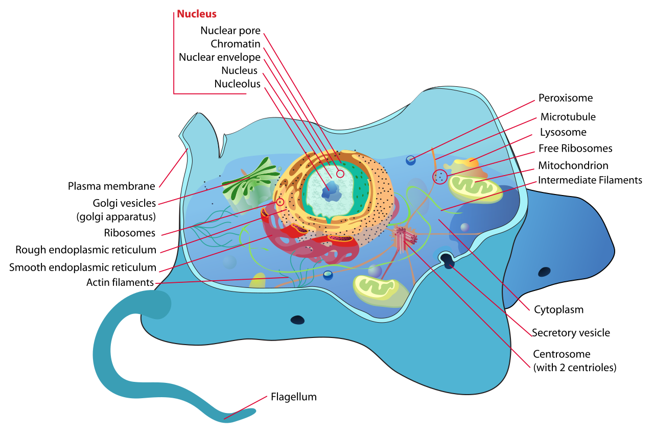
Animal Cell Structure
1.1.1 The cell as the unit of function #
A cell is a membrane-bounded compartment that can take up nutrients, convert energy, maintain internal conditions, and, given a suitable environment, grow and reproduce. Some organisms are one cell. Others are built by division and differentiation of many cells. In either case, the processes called “life” are cellular processes. An organism dies when its essential cells fail, and a tissue works only because its cells do specific jobs.

Cell Nucleus (Nih Bioart 72)
Cell biology asks three questions of every structure it meets. What is it made of? How is it arranged? What does that arrangement make possible that a bag of the same molecules could not do? The nucleus, a mitochondrion, a cilium, and a mitotic spindle are answers to that third question.
1.1.2 Scale #
Most bacteria are about 1–5 µm long. A typical animal cell is about 10–30 µm across, and a plant cell is often larger. A few specialised cells break these ranges: a human egg is about 100 µm, some neurons send axons for many centimetres, and the bacterium Thiomargarita can be visible to the naked eye.
These sizes set the methods. A light microscope can resolve about 0.2 µm, so it shows nuclei, mitochondria, and bacteria, but not ribosomes or a membrane bilayer. An electron microscope, or a fluorescence method that circumvents the diffraction limit, is required for those.
Useful reference sizes:
| Object | Approximate size |
|---|---|
| Small metabolite | < 1 nm |
| Globular protein | 3–10 nm |
| Plasma-membrane thickness | 5–8 nm |
| Ribosome | 25–30 nm |
| Virus (range is wide) | 20–300 nm |
| Bacterium | 1–5 µm |
| Mitochondrion | 0.5–1 µm wide, a few µm long |
| Nucleus of an animal cell | about 5–10 µm |
| Animal cell | about 10–30 µm |
Volume scales with the cube of linear size, and surface with the square. A larger cell has less surface per unit of cytoplasm, so diffusion and membrane area become limiting. Eukaryotes answer this partly by folding the plasma membrane and, more importantly, by adding internal membrane systems.
1.2 Cell theory #
The study of Cell theory provides fundamental principles governing cellular architecture, molecular interactions, and physiological regulation.

Cell Junction Simplified
1.2.1 How the theory was assembled #
The theory was not one experiment. It accumulated as lenses improved.
- Robert Hooke (1665) examined cork and named the empty compartments cells. He was looking at the walls of dead plant tissue, not at living contents.
- Antonie van Leeuwenhoek (1670s) ground single lenses of high quality and described living “animalcules” in water, including bacteria and protists. He was the first person to see living single cells.
- Robert Brown (1831) described the nucleus as a regular feature of plant cells.
- Matthias Schleiden (1838) concluded that plants are composed of cells and cell products. Theodor Schwann (1839) extended the claim to animals and argued that cells are the elementary particles of organisms. Together they stated the structural half of the theory.
- Rudolf Virchow (1855) supplied the generative half: omnis cellula e cellula, every cell from a cell. This rejected spontaneous generation of cells inside tissues.
Later work filled in mechanism rather than replacing the theory: chromosomes as the vehicles of heredity, DNA as the genetic material, and electron microscopy as the tool that showed the internal membranes Schleiden and Schwann could not have seen.
1.2.2 The statements, classical and modern #
The classical cell theory has three statements.
1. All living organisms are composed of one or more cells, and of material that cells have produced.
2. The cell is the basic unit of structure and function.
3. All cells arise from pre-existing cells by division.
A modern working version adds three further statements that every chapter below uses.
4. Cells of all organisms are similar in chemical composition and in the core reactions of metabolism.
5. Hereditary information passes from cell to cell through DNA, which is replicated and then partitioned.
6. Energy flows through cells. The immediate chemical currency is ATP, and the enzymes that make and spend it are cellular.
Viruses sit outside the theory. They have no metabolism of their own and replicate only inside a host cell. They are genetic packages that exploit cells, which is why cell biology studies them as tools and as parasites, not as a fourth kind of cell.
1.2.3 Two grades of organisation #
The deepest split visible with a microscope is the presence or absence of a nucleus.
- Prokaryotic cells (archaea and bacteria) have no nucleus and no mitochondria, chloroplasts, endoplasmic reticulum, or Golgi apparatus. Their DNA is a nucleoid. Ribosomes are 70S.
- Eukaryotic cells (plants, animals, fungi, protists) keep their chromosomes inside a nuclear envelope and divide the cytoplasm into membrane-bounded organelles. Ribosomes in the cytosol are 80S. Their mitochondria and chloroplasts retain 70S ribosomes, a trace of their bacterial origin.
“Prokaryote” is a grade, not a single evolutionary group. Archaea and bacteria are as distinct from each other, in membrane chemistry and in the machinery of transcription, as either is from eukaryotes. The three-domain scheme (Bacteria, Archaea, Eukarya) is the classification these notes use.
1.3 Prokaryotic cells #
The study of Prokaryotic cells provides fundamental principles governing cellular architecture, molecular interactions, and physiological regulation.
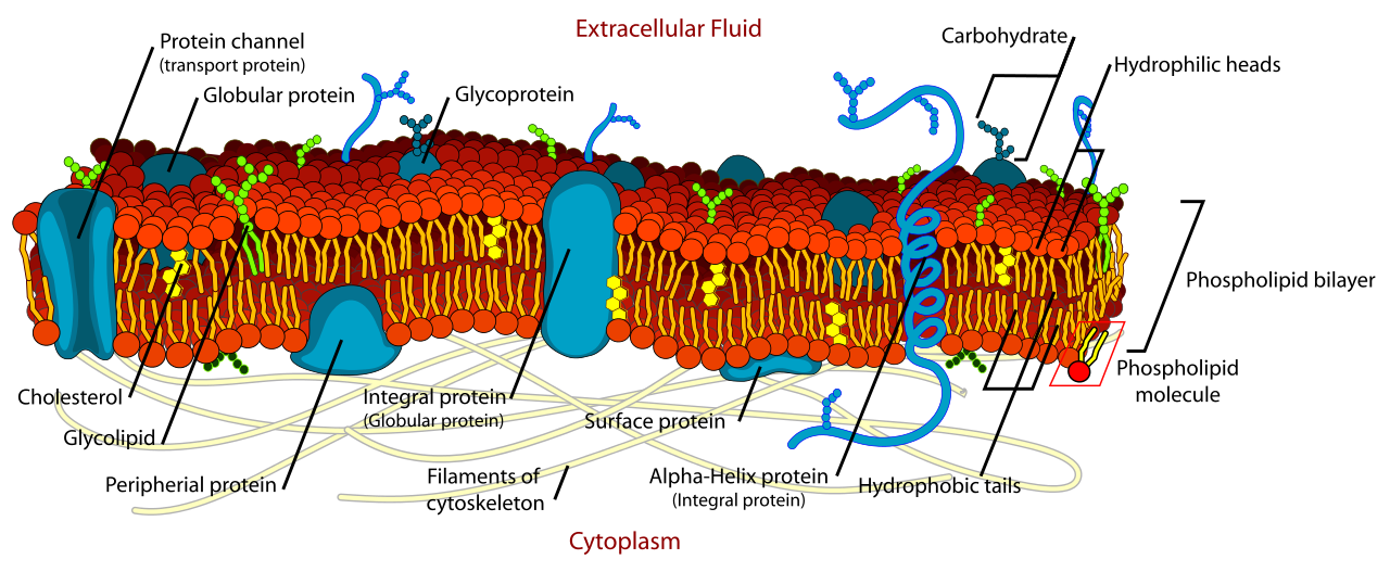
Cell Membrane Detailed
1.3.1 Shared plan #
A prokaryotic cell is not an empty sack. A typical cell has:
- a nucleoid, a region containing usually one circular chromosome of double-stranded DNA, compacted by supercoiling and nucleoid-associated proteins (not by histone octamers of the eukaryotic type);
- plasmids, smaller DNA circles that are optional and often carry accessory genes such as antibiotic resistance;
- 70S ribosomes (30S + 50S subunits), free in the cytoplasm;
- a plasma membrane, which is the site of energy metabolism because there are no mitochondria;
- a cell wall outside that membrane in most species;
- often a capsule, pili, or flagella.
There is no nucleolus and no mitosis. Division is binary fission: the chromosome is replicated, the two origins move apart, and the cell splits by ingrowth of the envelope, guided in bacteria by the FtsZ ring. Generation times can be as short as about 20 minutes under rich conditions, which is why bacteria dominate genetic experiments that need huge populations.
Older textbooks describe mesosomes, folded invaginations of the plasma membrane seen in chemically fixed bacteria, as the sites of respiration. In cells frozen rapidly rather than fixed with solvents, those folds largely disappear. They are fixation artifacts. Respiratory enzymes of bacteria sit in the plasma membrane itself.
1.3.2 Archaea #
Archaea are prokaryotic in cell plan and profoundly different in chemistry.
- Their membrane lipids use ether links between glycerol and long isoprenoid chains, not the ester-linked fatty acids of bacteria and eukaryotes. Tetraether lipids can span the membrane as a monolayer, which is stable at high temperature and extreme pH.
- Their walls do not contain peptidoglycan. Some have pseudopeptidoglycan (pseudomurein); others use protein or glycoprotein surface layers (S-layers).
- Their RNA polymerase and the proteins that handle DNA are closer to the eukaryotic machinery than to the bacterial one. This is a major reason Archaea are a separate domain, and a reason many researchers place eukaryotes as relatives of a specific archaeal group (Asgard archaea) rather than as a sister to all prokaryotes.
Archaea are not only extremophiles. They are abundant in ordinary soils and in the oceans, and methanogens among them are central to anaerobic carbon cycling.
1.3.3 Bacteria and the Gram envelope #
Bacteria (sometimes called eubacteria to distinguish them from archaea) have ester-linked fatty-acid membrane lipids and, with few exceptions such as Mycoplasma, a peptidoglycan wall. Peptidoglycan is a mesh of glycan strands, alternating N-acetylglucosamine and N-acetylmuramic acid, cross-linked by short peptides. The enzyme lysozyme cuts the glycan. Beta-lactam antibiotics block the peptide cross-links. Either treatment leaves the cell vulnerable to osmotic lysis, which is direct evidence that the wall, not the membrane, resists turgor.
The Gram stain reads the architecture of this envelope.
| Feature | Gram-positive | Gram-negative |
|---|---|---|
| Peptidoglycan | Thick, many layers | Thin |
| Outer membrane | Absent | Present, with lipopolysaccharide (LPS) on its outer face |
| Periplasm | Narrow | Distinct compartment between the two membranes |
| Teichoic acids | Present, woven through the wall | Absent |
| Gram stain | Purple; the thick wall retains the crystal-violet complex | Pink; alcohol extracts the complex, and the counterstain shows |
| Porins | Not applicable | Outer-membrane channels for small hydrophilic solutes |
LPS is a virulence factor. Its lipid A portion is the endotoxin that triggers strong inflammatory responses. The outer membrane is also a permeability barrier, which is why some antibiotics that kill Gram-positive species fail against Gram-negative ones.
Inside the cytoplasm, bacteria store reserves as inclusions: glycogen, polyphosphate, polyhydroxybutyrate, and, in cyanobacteria, protein-bounded carboxysomes that concentrate the carbon-fixing enzyme Rubisco. Magnetotactic bacteria contain magnetite crystals. These are compartments of a sort, but they are not homologous to eukaryotic organelles.
Bacterial flagella are rigid helical filaments of flagellin, rotated by a motor embedded in the envelope and powered by proton (or sometimes sodium) influx. They are unrelated, in structure and mechanism, to the microtubule-based flagella of eukaryotes. Sharing a name does not mean sharing a structure.
1.4 Eukaryotic cells #
The study of Eukaryotic cells provides fundamental principles governing cellular architecture, molecular interactions, and physiological regulation.

Cytokinesis Eukaryotic Mitosis
1.4.1 The compartment principle #
A eukaryotic cell keeps incompatible chemistries apart. Lysosomal hydrolases work at about pH 5; the cytosol is near pH 7.2. Oxidative phosphorylation needs a sealed inner mitochondrial membrane. Transcription and translation are separated in time and space: RNA is made and processed in the nucleus, and proteins are made in the cytosol. That separation is the structural reason eukaryotes can do regulated RNA splicing.
The price is traffic. Every protein made on cytosolic ribosomes has to be sent to the right compartment, and every time an organelle grows or divides, its membrane and its contents must be partitioned. Chapters 7 to 9 are about that logistics.
1.4.2 Animal and plant cells #
Both have a nucleus, endoplasmic reticulum, Golgi apparatus, mitochondria, peroxisomes, and a cytoskeleton. The differences that matter in a first course are these.

Desmosome Cell Junction
| Feature | Typical animal cell | Typical plant cell |
|---|---|---|
| Outer boundary | Plasma membrane plus extracellular matrix | Plasma membrane plus a rigid cell wall; neighbouring walls cemented by a pectin-rich middle lamella |
| Chloroplasts | Absent | Present in photosynthetic tissues; other plastids elsewhere |
| Vacuole | Small, several | Often one large central vacuole that stores solutes, maintains turgor, and can occupy most of the cell volume |
| Centrioles | Present in most animal cells; organise the spindle | Absent from flowering plants; the spindle is organised without centrioles |
| Cytokinesis | Contractile ring of actin and myosin pinches the cell in two | A cell plate, built from Golgi-derived vesicles, grows outward to make the new wall |
| Communication between cells | Gap junctions and other protein junctions | Plasmodesmata, cytoplasmic channels through the wall |
| Energy stores | Glycogen | Starch, inside plastids |
| Lysosomes | Prominent | The vacuole and lytic compartments do much of the same digestive work |
Fungal cells resemble animal cells in lacking chloroplasts, and plant cells in having walls. Fungal walls are chitin and glucans, not cellulose.
1.4.3 Three domains side by side #
| Feature | Bacteria | Archaea | Eukarya |
|---|---|---|---|
| Nucleus | No | No | Yes |
| Membrane lipids | Ester-linked fatty acids | Ether-linked isoprenoids | Ester-linked fatty acids |
| Peptidoglycan | Usually yes | No | No |
| Ribosomes | 70S | 70S | 80S in cytosol; 70S in mitochondria and chloroplasts |
| Chromosome | Usually one circular molecule | Usually circular | Multiple linear molecules, with telomeres, packaged by histones |
| Cell division | Binary fission, FtsZ | Binary fission | Mitosis and meiosis; cytokinesis by ring or cell plate |
| Organelles of the endomembrane system | No | No | Yes |
| Respiratory membrane | Plasma membrane | Plasma membrane | Inner mitochondrial membrane |
1.4.4 Endosymbiosis and why organelles have DNA #
Mitochondria descend from an alphaproteobacterium that took up residence inside an archaeal-related host cell. Chloroplasts descend from a later endosymbiosis with a cyanobacterium. The evidence is convergent:

Trad Chloroplast Primary Endosymbiosis
- both organelles have a double membrane;
- both contain a circular chromosome and 70S ribosomes;
- both divide by fission, and a cell cannot build them from scratch;
- their gene sequences group with bacteria, not with the nuclear genome.
Most of the original bacterial genes have moved to the nucleus. The organelle now imports the great majority of its proteins from the cytosol (Chapter 9). What remains in the organelle genome is a short list of hydrophobic core subunits of the energy-converting membranes, plus the RNAs needed to translate them. Human mitochondrial DNA is a circle of 16,569 base pairs and carries 37 genes: 13 proteins, 22 transfer RNAs, and 2 ribosomal RNAs. In animals it is inherited almost entirely from the egg, because sperm mitochondria are few and are usually eliminated after fertilisation.
1.5 Cells as experimental models #
The study of Cells as experimental models provides fundamental principles governing cellular architecture, molecular interactions, and physiological regulation.

Human Cell Nucleus Numbered Version
1.5.1 Why models, and which ones #
Cell processes are conserved enough that a mechanism found in a yeast often holds in a human cell. Models are chosen because a question is easier to ask in them: short generation time, transparent body, giant cells, or genetics that are already done.
| Model | What it is good for |
|---|---|
| Escherichia coli | Prokaryotic molecular genetics, gene regulation, recombinant DNA |
| Budding yeast Saccharomyces cerevisiae | Eukaryotic cell cycle, secretion, and genetic screens. Many cdc genes were found here |
| Fission yeast Schizosaccharomyces pombe | Cell-cycle control, especially the CDK that Nurse identified as Cdc2 |
| Caenorhabditis elegans | The complete cell lineage of an animal, and the genetics of apoptosis |
| Drosophila melanogaster | Developmental patterning, chromosome biology |
| Arabidopsis thaliana | Plant cell biology, a small genome among flowering plants |
| Xenopus laevis eggs | Cell-free extracts for the cell cycle; the egg is large enough to inject |
| Mouse, and human or mouse cells in culture | Mammalian signalling, development, and disease mutations |
| HeLa and other immortal lines | Biochemistry that needs grams of uniform human cells |
A cultured line is a tool with a history. HeLa cells, established in 1951 from Henrietta Lacks’s cervical tumour without her consent, grow so well that they have contaminated many other lines. A result attributed to “human cells” is only as good as the authentication of the line.
1.5.2 In vitro, in culture, and in the organism #
Three levels of experiment answer different questions.
- A cell-free extract (cytosol plus added organelles, or a purified protein) asks whether a process can run without the intact cell. Nuclear import, ER translocation, and the cell cycle have all been reconstituted this way.
- Cell culture asks what one cell type does when hormones, drugs, or genetic changes are applied under controlled conditions. Primary cultures retain normal biology for a limited number of divisions. Immortal lines are convenient and genetically abnormal.
- The intact organism asks whether the mechanism matters to development, physiology, or disease. A gene that is essential in culture may be redundant in a mouse, and the reverse also happens.
Good cell biology moves between these levels instead of trusting only one.
1.6 Key terms, summary, and self-check #
Key terms. Cell theory; prokaryote; eukaryote; domain; nucleoid; peptidoglycan; Gram-positive; Gram-negative; LPS; endosymbiosis; model organism.
Summary. Cells are the units of structure, function, and descent. Archaea and bacteria lack a nucleus and differ from each other in membrane and wall chemistry. Eukaryotes compartmentalise the cytoplasm, and their mitochondria and chloroplasts are descended from bacteria. Experimental cell biology concentrates on a few species because the core machinery is shared.
Self-check.
1. Which half of the cell theory belongs to Virchow, and what observation forced it?
2. Why does the Gram stain come out different colours? Answer in terms of envelope structure, not in terms of the dye recipe alone.
3. Give four independent lines of evidence that mitochondria came from bacteria.
4. Why is a mesosome no longer accepted as a respiratory organelle?
5. A student says yeast results are irrelevant to human cells. Give one historical counterexample from the cell cycle, and one reason the claim sometimes is true.
