
5.1 Making ions #
5.1.1 Soft ionisation #
A mass spectrometer manipulates ions in electric or magnetic fields. Neutral molecules are invisible to it, and hard ionisation shatters a protein. Fenn developed electrospray of macromolecules. MALDI was developed by Karas and Hillenkamp and, in the form recognised with Fenn, by Tanaka.
MALDI embeds the analyte in a large excess of a small molecule that absorbs the laser, isolates analyte molecules from each other, and provides the charge. The usual product is singly charged, so m/z is essentially the mass. A 100 kDa protein at charge +1 therefore needs an analyser that can handle m/z 100 000. Time-of-flight can. The matrix crystal is not a native environment: MALDI is a mass method, not a fold method.
Electrospray pushes solution through a charged needle. Droplets shrink by evaporation and fission until gas-phase ions remain. Proteins pick up many protons in the usual positive-ion experiment; nucleic acids are often run as negative ions. The spray is soft enough that non-covalent complexes can survive, and it couples naturally to liquid chromatography.
5.1.2 The charge-state envelope #
The analyser reports m/z, not mass. A protein of mass M daltons carrying z protons appears near
m/z = (M + z) / z
One pure protein therefore gives a ladder of peaks, a charge-state envelope. If m_z and m_{z+1} are adjacent peaks, z = (m_{z+1} − 1) / (m_z − m_{z+1}), and M follows. Deconvolution does this for the whole ladder and returns a zero-charge mass. An intact mass accurate to a few daltons shows a missing terminal residue or a modification of known mass, such as a phosphate of about 80 Da. It does not sequence the chain: same composition means same mass. Sodium or potassium adducts shift and split peaks and can be mistaken for modifications. Unfolded chains typically carry more charge than folded ones because more basic sites are exposed, so the envelope’s position is a rough report of compactness in the ion that left the droplet, not a structure.
5.2 Separating ions and fragmenting them #
5.2.1 Quadrupole, time-of-flight, Orbitrap #
Resolution m/Δm is the mass divided by the smallest separable difference. Mass accuracy is how close the measured m/z lies to the true value. High resolution can still be inaccurate if the calibration is wrong.
| Analyser | What sorts the ions | Consequence |
|---|---|---|
| Quadrupole | a stable trajectory in an oscillating field between four rods | one m/z window passes. Modest resolution. The selector in tandem instruments |
| Time-of-flight | flight time after acceleration. With ½ mv² = z e V, m/z is proportional to t² | wide mass range; the partner of MALDI. A reflectron corrects the energy spread |
| Orbitrap | oscillation frequency about a central electrode, proportional to 1/√(m/z), read as an image current and Fourier-transformed | high resolution without a superconducting magnet |
| FT-ICR | cyclotron frequency ω = z e B / m | the highest resolution; needs a strong magnet |
A quadrupole in front of a time-of-flight or an Orbitrap selects a precursor, a cell fragments it, and the second analyser reads the pieces.
5.2.2 Tandem mass spectrometry #
Peptides in collision-induced dissociation break mainly at the peptide bond. A b ion keeps the N-terminus. A y ion keeps the C-terminus. A ladder of either differs by residue masses, which is how a sequence is read. Leucine and isoleucine have the same mass. Glutamine and lysine differ by about 0.036 Da and separate only if the accuracy is good enough. A labile modification can leave as a neutral during collision, so the fragments look unmodified. Electron-transfer dissociation breaks the backbone differently and more often keeps such modifications. The fragmentation method changes what chemistry survives. The spectrometer still measures m/z.
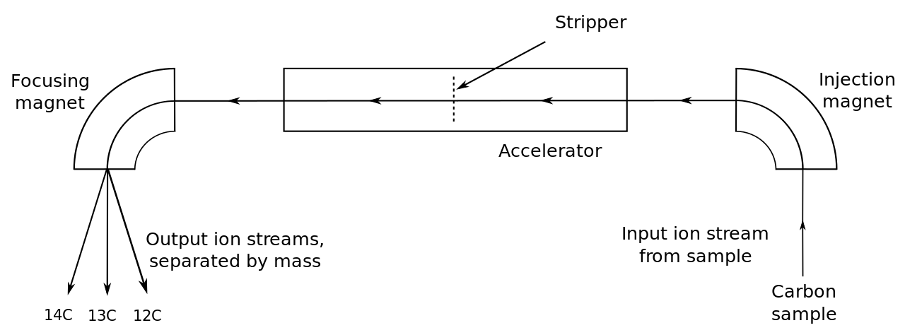
Accelerator Mass Spectrometer Schematic For Radiocarbon
5.2.3 Proteomics as a data path #
The data path, not a bench protocol, has four stages. Proteins are digested, usually with trypsin, which cuts on the carboxyl side of lysine and arginine unless proline follows, giving peptides of a useful size with a basic C-terminus. Peptides are separated by reversed-phase chromatography because the instrument cannot sequence hundreds at once; it selects ions as they elute, or fragments broader windows. Each precursor is fragmented. Software then scores the fragment spectrum against peptides predicted from a sequence database. A peptide-spectrum match is a hypothesis with a score.
Random matches score well occasionally. A decoy database of reversed or shuffled sequences estimates how often. Identifications are reported at a stated false-discovery rate, the expected fraction of the accepted list that is chance. A rate near 1% is a common standard, not a law. One weak peptide is poorer evidence than several independent peptides. Data-dependent acquisition spends its time on the strongest ions, and cellular abundances span many orders of magnitude, so absence from the list is not absence from the cell. A heavy-isotope partner of the same peptide gives a relative amount. An absolute concentration needs a standard of known quantity. Scoring algorithms sit with Bioinformatics. The defendable statement is that an ion at a measured m/z fragmented to a measured pattern and survived a decoy test.
5.2.4 Native mass spectrometry and hydrogen–deuterium exchange #
Native MS electrosprays complexes from a volatile salt solution so that non-volatile components such as phosphate do not destroy them or swamp the spectrum. The mass is the mass of what survived. Four times the subunit mass suggests a tetramer if the subunits are identical and nothing else is bound. A complex that disappears may have been real in solution and too fragile for the spray. Stoichiometry is a mass result. Subunit arrangement is Structural Biology.

Mass Spectrometer Schematic-Af
Hydrogen–deuterium exchange uses mass differently. Backbone amides exchange with solvent deuterium more slowly when hydrogen-bonded or buried, and the protein gains about 1 Da per site exchanged. Peptide-level spectra after the exchange has been slowed map which stretches were exposed. Exchange is acid- and base-catalysed and slowest near pH 2.5 in the cold, which is why a difference can be preserved long enough to measure. Back-exchange, loss of deuterium before detection, makes the measured uptake an underestimate. The pattern reports protection and opening. It is not itself a structure.
5.3 Key terms, summary, and self-check #
Key terms. Electrospray; MALDI; m/z; charge-state envelope; quadrupole; time-of-flight; Orbitrap; b ion; y ion; decoy database; false-discovery rate; native MS; hydrogen–deuterium exchange.
Summary. Soft ionisation puts proteins into the gas phase intact enough to weigh. Electrospray gives a charge-state envelope whose spacing yields the mass; MALDI usually gives a single charge and needs a wide mass range. Tandem MS reads peptide ladders, with hard ambiguities where residues share a mass. A database match needs a false-discovery rate. Native MS adds stoichiometry of surviving complexes, and hydrogen–deuterium exchange adds a map of amide protection.
Self-check.
1. Why does one pure protein give many electrospray peaks, and how do two adjacent peaks yield the mass?
2. What demand does a singly charged 100 kDa MALDI ion place on the analyser, compared with an electrospray envelope of the same protein?
3. What does a y ion contain that a b ion does not, and why can leucine not be told from isoleucine by mass alone?
4. What does a decoy database estimate, and why is “not on the list” not “not in the cell”?
5. Native MS shows four times the subunit mass. What stoichiometry does that suggest, and why might a genuine complex still be missing?
