
9.1 Evolution of Nitrogenous Waste Excretion #
Catabolism of amino acids and nucleic acids generates toxic ammonia (NH3). Vertebrates have evolved three primary chemical strategies for detoxifying and eliminating nitrogenous wastes:
                          PROTEINS & NUCLEIC ACIDS
                                      │
                                      ▼
                                AMMONIA (NH3)
                                      │
        ┌─────────────────────────────┼─────────────────────────────┐
        ▼                             ▼                             ▼
   AMMONOTELISM                  UREOTELISM                    URICOTELISM
 (Aquatic Teleosts)         (Mammals, Amphibians)          (Birds, Reptiles)
Toxic, high water loss      Low toxicity, urea cycle       Insoluble paste, zero water
1. Ammonotelism (Ammonia Excretion): Highly toxic, alkaline, and membrane-permeable. Requires vast amounts of water for dilute excretion (>500 mL H2O/g N). Utilized by freshwater bony fishes, larval amphibians, and aquatic invertebrates that vent ammonia directly across gill membranes via simple diffusion.
2. Ureotelism (Urea Excretion): Ammonia is converted into water-soluble, non-toxic urea via the hepatic ornithine-urea cycle at an energetic cost of 4–5 ATP equivalents per urea molecule. Urea requires modest water for excretion (50 mL H2O/g N). Utilized by adult amphibians, mammals, and chondrichthyan fishes (sharks retain urea and TMAO to maintain hyperosmotic blood).
3. Uricotelism (Uric Acid Excretion): Complex, energetically expensive purine pathway converting ammonia into uric acid and guanine. Highly non-toxic and virtually insoluble in water; precipitates as a crystalline white semi-solid paste requiring minimal water (<5 mL H2O/g N). Indispensable evolutionary adaptation for animals enclosed in cleidoic (shelled) eggs (birds, reptiles, insects) where toxic soluble wastes would poison the developing embryo.
9.2 Osmoregulation in Aquatic Environments #
The study of Osmoregulation in Aquatic Environments provides fundamental principles governing cellular architecture, molecular interactions, and physiological regulation.
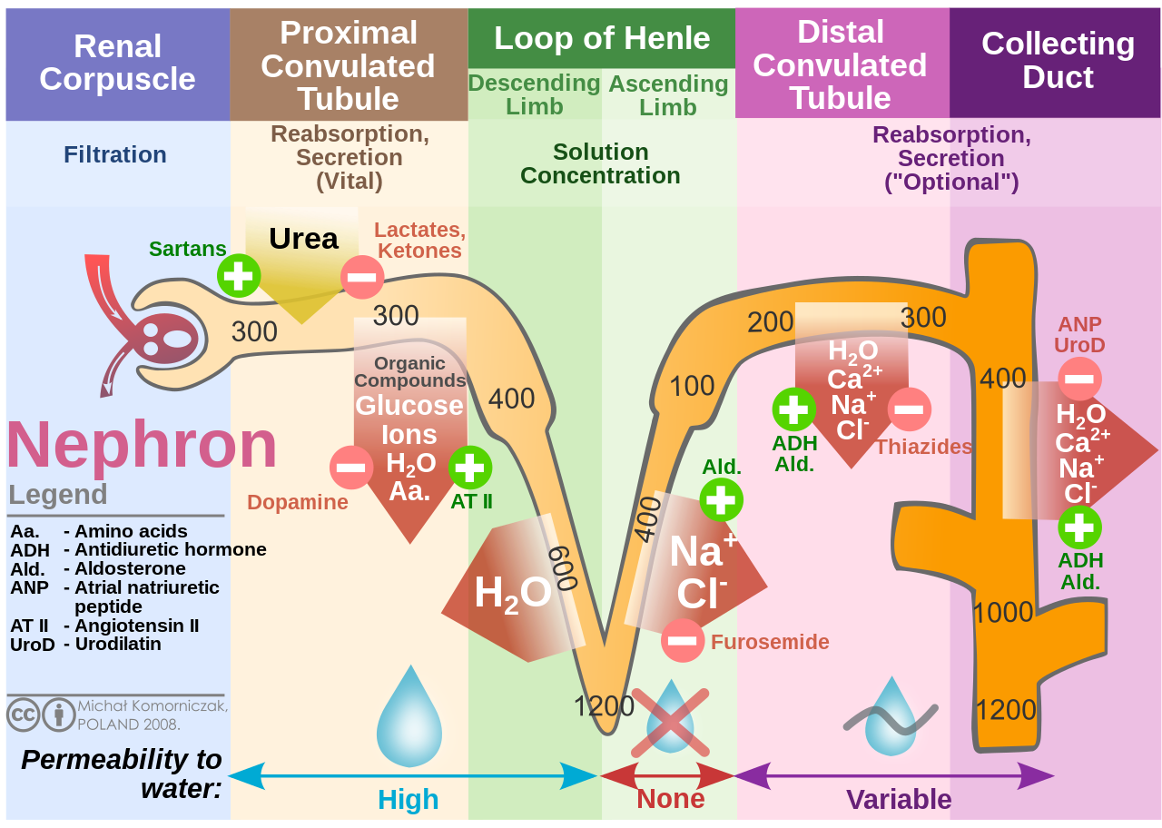
Kidney Nephron Molar Transport
9.2.1 Freshwater Bony Fishes (Hyperosmotic Regulators) #
- Challenge: Body fluids are hyperosmotic to freshwater (300 mOsm/L vs. 5–10 mOsm/L). Water floods into the fish osmotically across gills and oral mucosa; salts are lost continuously via passive diffusion.
- Adaptations:
1. Never drink water.
2. Large, highly developed renal glomeruli producing vast quantities of extremely dilute, hypoosmotic urine.
3. Specialized chloride cells (mitochondria-rich cells) in gill epithelium actively pump sodium (Na+) and chloride (Cl–) from dilute ambient water into the blood against steep concentration gradients using apical Na+/H+ and Cl–/HCO3– antiporters powered by basolateral Na+/K+-ATPases.
9.2.2 Marine Bony Fishes (Hypoosmotic Regulators) #
- Challenge: Body fluids are hypoosmotic to seawater (300 mOsm/L vs. 1,000 mOsm/L). Water is continuously drawn out of the fish by osmosis; salts diffuse continuously into the blood.
- Adaptations:
1. Drink seawater continuously (10–20% of body weight daily) to replace osmotic water loss.
2. Ingested seawater is absorbed across the intestine; excess divalent ions (Mg2+, SO42-) are voided in feces.
3. Reduced or absent glomeruli (aglomerular kidneys in pipefish, toadfish) produce tiny volumes of concentrated isosmotic urine, minimizing urinary water loss.
4. Specialized chloride cells in gills reverse direction, actively pumping excess Na+ and Cl– out of the blood directly into the hyperosmotic sea.
9.3 The Mammalian Juxtamedullary Nephron #
The mammalian kidney consists of an outer cortex and an inner renal medulla. The functional unit is the nephron:
1. Glomerulus & Bowman’s Capsule: Blood enters via the wide afferent arteriole and exits via the narrower efferent arteriole, generating high hydrostatic filtration pressure (55 mmHg). Glomerular capillary endothelial fenestrations, the basement membrane, and podocyte slit diaphragms form a molecular sieve filtering water, ions, glucose, and urea, while excluding blood cells and large proteins (Glomerular Filtration Rate, GFR ≈ 125 mL/min or 180 L/day).
2. Proximal Convoluted Tubule (PCT): Lined by simple cuboidal epithelium with a dense brush border of microvilli. Reabsorbs 65–70% of the filtered water and Na+, and 100% of filtered glucose and amino acids via secondary active transport coupled to Na+-glucose cotransporters (SGLT1/2).
3. Loop of Henle (Countercurrent Multiplier):
- Descending Thin Limb: Highly permeable to water via constitutive Aquaporin-1 (AQP1) channels; impermeable to NaCl and urea. Water flows osmotically into the hypertonic medullary interstitium, concentrating tubular fluid up to 1,200 mOsm/L at the hairpin turn.
- Ascending Thick Limb (TAL): Completely impermeable to water. Features apical Na+-K+-2Cl– cotransporters (NKCC2)* actively pumping ions out of the tubular fluid into the medullary interstitium. This dilutes the luminal fluid down to 100 mOsm/L (“diluting segment”) while building the hyperosmotic medullary gradient.
4. Vasa Recta (Countercurrent Exchanger): Sluggish, hairpin capillary loops mirroring the loop of Henle that supply oxygen to the medulla while passively exchanging water and solutes, preventing the washout of the hyperosmotic medullary salt gradient.
9.4 Endocrine Regulation of Renal Function #
Renal conservation of water and blood pressure homeostasis is governed by two master endocrine pathways:
9.4.1 The Renin-Angiotensin-Aldosterone System (RAAS) #
1. Declining blood pressure, renal sympathetic activation, or low NaCl delivery to the macula densa triggers the juxtaglomerular (JG) cells to secrete the aspartyl protease renin into the circulation.
2. Renin cleaves circulating liver-derived angiotensinogen into the decapeptide angiotensin I (Ang I).
3. Pulmonary capillary Angiotensin Converting Enzyme (ACE) cleaves Ang I into the active octapeptide Angiotensin II (Ang II).
4. Ang II exerts powerful physiological homeostatic responses:
- Systemic vasoconstriction, raising total peripheral resistance.
- Stimulates thirst in the hypothalamus.
- Directs the adrenal cortex (zona glomerulosa) to secrete Aldosterone.
- Aldosterone up-regulates apical ENaC sodium channels and basolateral Na+/K+-ATPases in cortical collecting duct principal cells, driving active Na+ reabsorption and potassium (K+) excretion.
9.4.2 Antidiuretic Hormone (ADH / Vasopressin) #
1. Dehydration and hyperosmolar plasma (>285 mOsm/kg) are detected by anterior hypothalamic osmoreceptors, triggering the posterior pituitary (neurohypophysis) to release Antidiuretic Hormone (ADH).
2. ADH binds to V2 basolateral receptors on collecting duct principal cells, activating adenylate cyclase to produce cyclic AMP (cAMP) and stimulate Protein Kinase A (PKA).
3. PKA phosphorylates intracellular vesicles containing Aquaporin-2 (AQP2) water channels, triggering their rapid exocytic insertion into the apical luminal membrane.
4. Water is drawn osmotically out of the collecting duct lumen across AQP2 channels into the hyperosmotic medullary interstitium, concentrating urine up to a maximum of 1,200–1,400 mOsm/L in humans (and >5,000 mOsm/L in desert kangaroo rats, Dipodomys).
9.5 Key Takeaways & Exam High-Yield Summary #
- Loop of Henle Length Dictates Urine Concentration: Desert mammals (Dipodomys) possess exceptionally long loops of Henle extending deep into thick renal medullas, generating extreme hyperosmotic medullary gradients; aquatic beavers possess short cortical nephrons.
- NKCC2 Target of Loop Diuretics: Furosemide (Lasix) inhibits the Na+-K+-2Cl– cotransporter in the thick ascending limb, abolishing the medullary gradient and inducing massive diuresis.
- Marine Teleosts vs. Sharks: Marine teleosts drink seawater and excrete salt across gills; marine sharks retain high concentrations of urea and trimethylamine oxide (TMAO) in blood, making them slightly hyperosmotic to seawater (preventing water loss).
9.6 Review & Self-Assessment Questions #
1. Explain the molecular etiology and clinical manifestations of Central vs. Nephrogenic Diabetes Insipidus.
- Answer: Central Diabetes Insipidus results from failure of the posterior pituitary to synthesize or secrete ADH (due to head trauma or hypothalamic tumors). Nephrogenic Diabetes Insipidus results from mutated, defective renal V2 receptors or non-functional AQP2 channels in the collecting duct. Both present with severe polyuria (>10 L/day of dilute urine) and compensatory polydipsia.
2. Why does the thick ascending limb of the loop of Henle require high mitochondrial density?
- Answer: The thick ascending limb actively pumps Na+, K+, and Cl– ions against steep electrochemical gradients via NKCC2 cotransporters and basolateral Na+/K+-ATPases, demanding vast amounts of ATP generated by oxidative phosphorylation.
—
—
—