
8.1 Tube, crest, and migration #
8.1.1 Neural tube and neural crest #
The nervous system begins as ectoderm. Underlying mesoderm, and the organiser, induce the neural plate rather than skin. The molecules of that decision, including bone morphogenetic protein and its inhibitors, are Developmental Biology and are not repeated. The plate folds into a neural tube, closing from the middle toward both neuropores. The cavity becomes the ventricles. The wall becomes the central nervous system, retina included. Failure of closure leaves brain or cord open to the surface.
Cells at the crest of the tube migrate as the neural crest and become dorsal-root and autonomic neurons, Schwann cells, enteric neurons, and also melanocytes and the adrenal medulla.
8.1.2 Proliferation, migration, and differentiation #
Progenitors divide next to the ventricle. Early on they are radial glia, both the stem cells and the scaffold from ventricle to pia. Cortical pyramidal cells migrate along that scaffold in inside-out order: deep layers settle first, and later cells pass them.
Sonic hedgehog from the floor plate ventralises the tube, so motor neurons are born ventrally, while roof-plate signals dorsalise sensory territories. Inhibitory interneurons of the cortex are born ventrally, in the ganglionic eminences, and migrate in from the side. Glutamate and GABA identities are already biased before the cells meet. Transcription factors set transmitter, channels, and shape. Activity later edits connections.
8.2 Guidance and the first synapse #
The study of Guidance and the first synapse provides fundamental principles governing cellular architecture, molecular interactions, and physiological regulation.
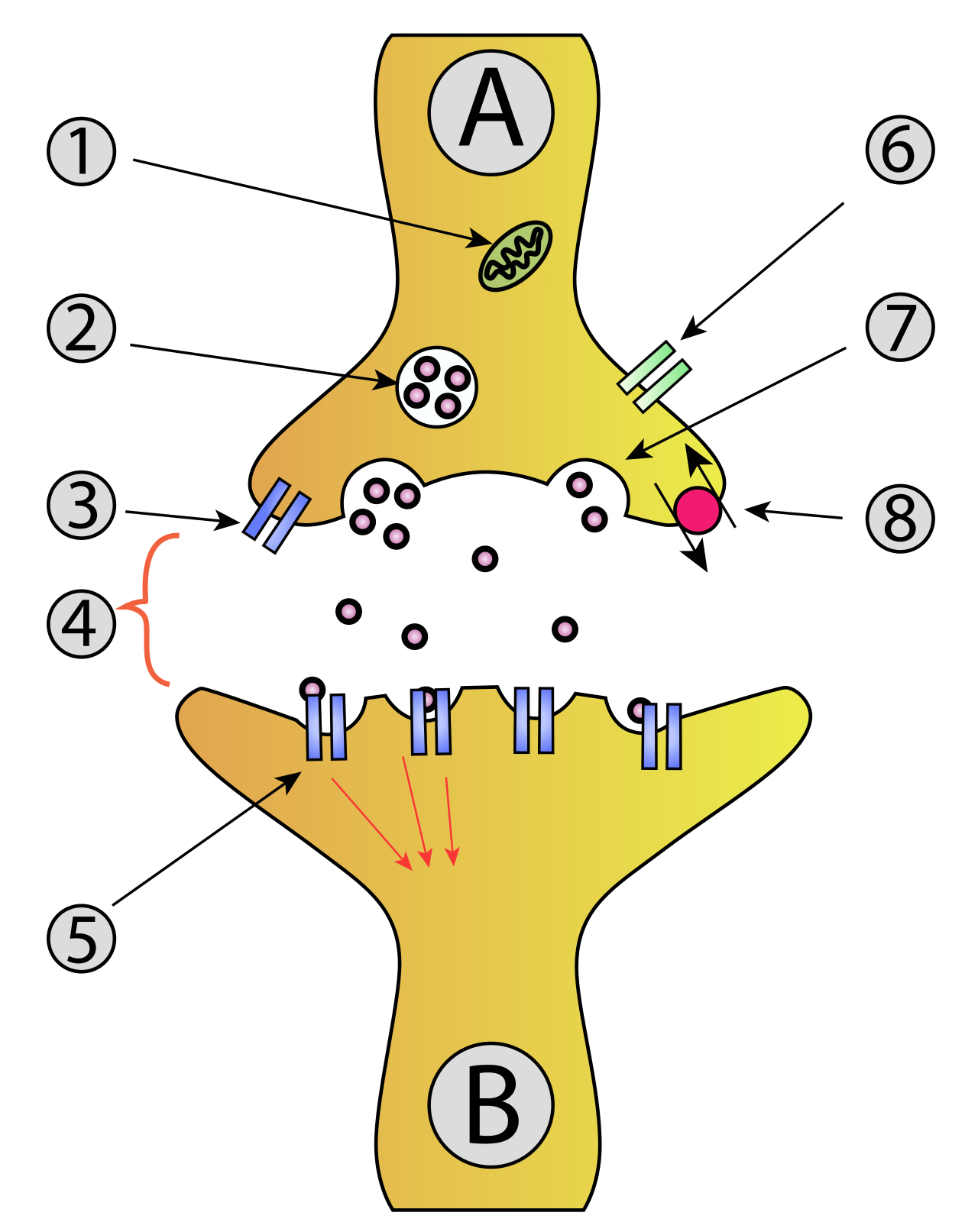
Synapse Diag1
8.2.1 Growth cones and four cue families #
A growth cone is a motile tip of actin filopodia and invading microtubules, the crawling machinery of Cell Biology aimed by receptors. A cue is not attractive or repulsive in itself. The receptor decides the sign.
| Family | Typical receptor | Classical sign | Example |
|---|---|---|---|
| Netrins | DCC attracts; Unc5 repels | Either | Floor plate draws commissural axons |
| Slits | Robo | Repulsive | Stops an axon recrossing the midline |
| Semaphorins | Plexin, neuropilin | Mostly repulsive | Keeps axons out of the wrong territory |
| Ephrins | Eph receptors | Often repulsive in maps | Gradients that set topography |
Commissural axons are drawn toward the floor plate by netrin acting on DCC. After they cross, Robo makes them sensitive to Slit, and Slit repels them, so they turn along the cord and do not cross back. Sperry rotated a frog’s eye and cut the optic nerve. Regenerating axons returned to their old tectal addresses, and the frog struck as if the world had rotated with the eye. They had followed labels, not success. In that map, temporal axons rich in EphA receptors are repelled by posterior ephrin-A, and nasal axons tolerate the same gradient.
8.2.2 Forming a synapse, then keeping it #
Contact is not yet a synapse. At the neuromuscular junction the axon releases agrin, which with LRP4 activates muscle MuSK. Through rapsyn, nicotinic receptors cluster under the terminal, acetylcholinesterase occupies the basal lamina, and receptors elsewhere on the fibre disappear. The adult ε subunit replaces the fetal γ subunit. Central synapses have no single agrin; neurexins and neuroligins are one adhesion pair among several.
Early wiring is excessive. Neurons that fail to capture a target-derived neurotrophin, carried home by retrograde transport, are removed by apoptosis, using the death machinery of Cell Biology. Axons from the two eyes first overlap in thalamus and cortex, then segregate.
8.3 Critical periods #
8.3.1 Ocular dominance, and what closes the window #
Axons that fire together keep their synapses on the same cells. Axons out of step lose them. The two eyes are rarely correlated, so their terminals separate into eye-specific geniculate layers and into ocular dominance columns in V1. Waves of spontaneous retinal activity begin the sorting before vision. Hubel and Wiesel then closed one eye of a young animal. Cortical cells shifted to the open eye, and the closed eye’s columns shrank, although the retina still worked. The same closure in an adult did little. The window is a critical period. Closing both eyes disturbs the balance between the eyes less than closing one, because the eyes compete for territory and the loss is not mere disuse.
Adult synapses do not freeze when the period ends. What ends is the capture of whole territories. Maturing inhibition, especially from parvalbumin interneurons, is required for the window to open on time and is associated with its close. Perineuronal nets build up around those cells and restrict further structural change. Myelin inhibits sprouting.
8.4 Key terms, summary, and self-check #
Key terms. Neural tube; neural crest; radial glia; inside-out migration; growth cone; netrin; slit; semaphorin; ephrin; agrin; ocular dominance; critical period; monocular deprivation.
Summary. The neural tube forms the central nervous system and the neural crest forms most peripheral neurons and Schwann cells, after inductions taught in Developmental Biology. Pyramidal cells migrate inside-out on radial glia; cortical interneurons migrate in from the ventral forebrain. Growth cones read netrins, slits, semaphorins, and ephrins according to their receptors. Synapses are induced by clustering signals, clearest for agrin at the neuromuscular junction, then pruned by neurotrophins and by activity. Monocular deprivation during a critical period lets the open eye take cortical territory. The period closes as inhibition and the extracellular matrix mature.
Self-check.
1. Which peripheral neurons are neural crest rather than neural tube?
2. Why do later-born cortical neurons settle further from the ventricle?
3. After a commissural axon has crossed, which cue family repels it from the midline?
4. In Sperry’s rotated eye, why did regenerating axons ignore the eye’s new orientation?
5. Why is closing one eye worse for ocular balance than closing both for the same time?
—
—
—