
 SVG
SVGAction Potential Basic Shape
Basic shape of an action potential, with all complicating elements omitted, showing only labels for time and membrane potential
 SVG
SVGAll Or None Law
This detailed scientific diagram illustrates All Or None Law in the context of Neurobiology.
 SVG
SVGAxo Axonic Synapse
Neuronal electrical signaling relies on rapid, transient voltage reversals known as action potentials propagating along excitable axolemmas.
 SVG
SVGShowing The Lobes Of The Brain
This detailed scientific diagram illustrates Showing The Lobes Of The Brain in the context of Neurobiology.
 SVG
SVGImgnotraçat Arc Reflex Eng
This image was provided to Wikimedia Commons as a contribution from an art & design school thanks to a collaboration between Serra i Abella and Amical Wikimedia.
 SVG
SVGNeuromuscular Junction
Diagram representing a neuromuscular junction, showing ACh receptors and mitochondria of the neuron.
 SVG
SVGNeuron
Neuronal electrical signaling relies on rapid, transient voltage reversals known as action potentials propagating along excitable axolemmas.
 SVG
SVGPropagation Of Action Potential Along Myelinated Nerve Fiber
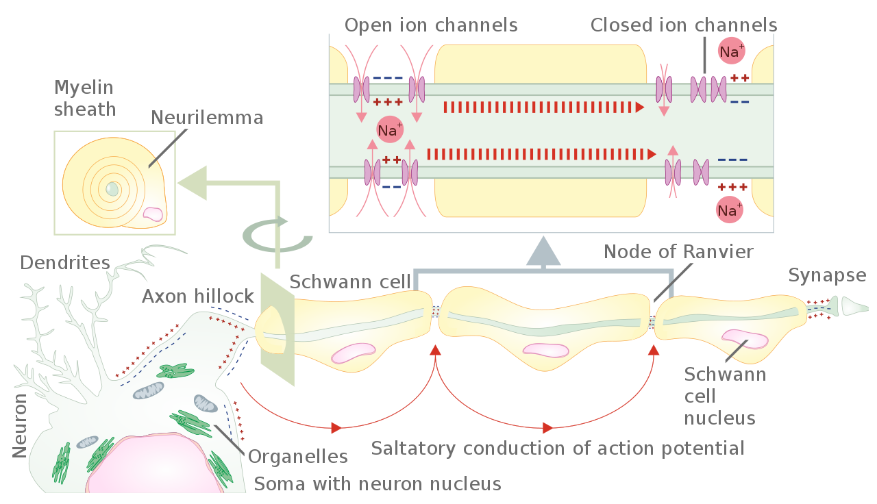
Translation and vector of the File:Propagation of action potential along myelinated nerve fiber.png Image description: Schematic representation of the action potential propagation through myelinated nerve fiber of peripheral nervous system. From axon hillock of neuron body (soma) action potential propagates from one unmyelinated fiber part to the next one. The unmyelinated parts of the nerve fiber
 SVG
SVGSynapse Diag1
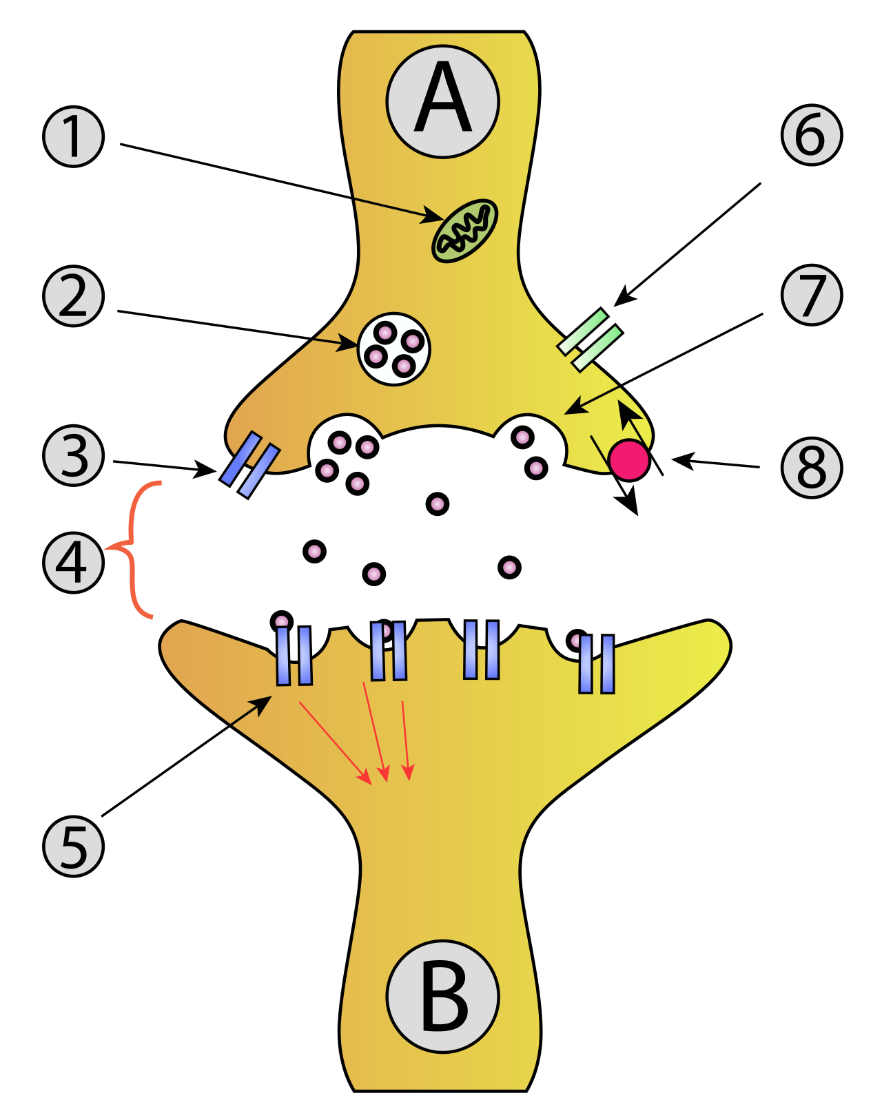
Synaptical transmission (chemical). A: Neuron (Presynaptic) B: Neuron (Postsynaptic) Mitochondria Synaptic vesicle full of neurotransmitter Autoreceptor Synaptic cleft Neurotransmitter receptor Calcium Channel Fused vesicle releasing neurotransmitter Neurotransmitter re-uptake pump
 SVG
SVGNeuron, Langneutral
Neuronal electrical signaling relies on rapid, transient voltage reversals known as action potentials propagating along excitable axolemmas.
 SVG
SVGNeuron Hand-Tuned2
Neuronal electrical signaling relies on rapid, transient voltage reversals known as action potentials propagating along excitable axolemmas.
 PNG
PNGAction Potential Vert
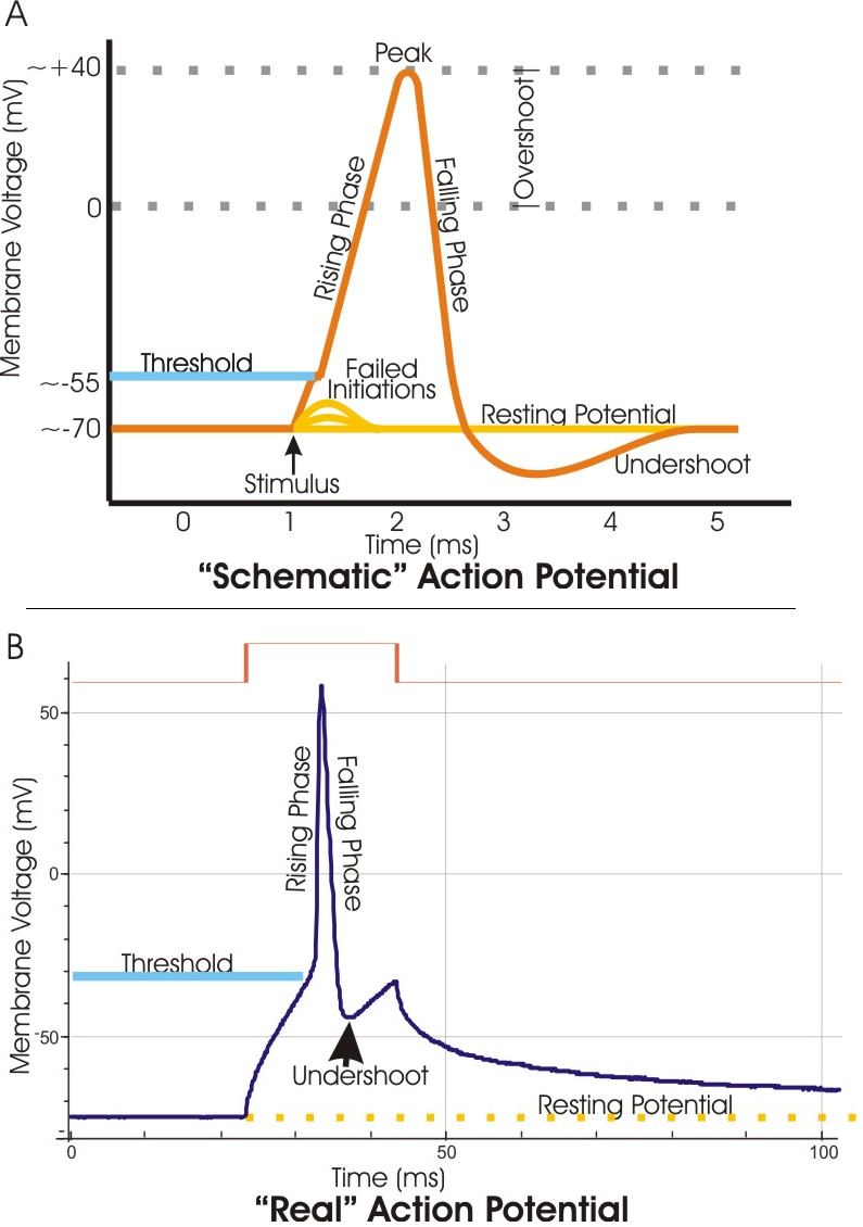
A. Schematic of an electrophysiological recording of an action potential showing the various phases which occur as the wave passes a point on a cell membrane . B. An actual action potential (blue trace) recorded from a mouse hippocampal pyramidal neuron. In this case, the action potential was stimulated by a prolonged pulse of current (brown trace; approx. 2 micro Amps) passed into the cell through the recording electrode. This method of stimulation distorts the AP compared to the schematic, in that the "real" action potential is sitting atop a voltage offset caused by the current pulse. Thus, for example, the "undershoot" is offset from the resting potential , although it would dip below rest if the offset were not present. The slow decline of the membrane potential back toward rest upon the termination of the current pulse reflects the long time constant of the neuronal membrane.
 PNG
PNGSpike Clusters
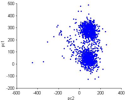
2D projections of the weight of the 1st (y-axis) and 2nd (x-axis) principle component of neuronal spikes shapes. Each cluster presumably collates spikes (see action potential ) of one neuron. This type of projection is used in spike sorting .
 PNG
PNGGehînke Ku
Communication at chemical synapses requires release of neurotransmitters. When the presynaptic membrane is depolarized, voltage-gated Ca 2+ channels open and allow Ca 2+ to enter the cell. The calcium entry causes synaptic vesicles to fuse with the membrane and release neurotransmitter molecules into the synaptic cleft. The neurotransmitter diffuses across the synaptic cleft and binds to ligand-gated ion channels in the postsynaptic membrane, resulting in a localized depolarization or hyperpolarization of the postsynaptic neuron
 PNG
PNGNeuro Muscular Junction
A neuromuscular junction (or myoneural junction) is a chemical synapse formed by the contact between a motor neuron and a muscle fiber. It is at the neuromuscular junction that a motor neuron is able to transmit a signal to the muscle fiber, causing muscle contraction. Labels from top right: myelin sheath , mitochondria , muscle fiber , ach receptors (acetylcholine), neurotransmitters , schwann cell , axoplasm , neurilemma .
 PNG
PNGNodes Of Ranvier
This detailed scientific diagram illustrates Nodes Of Ranvier in the context of Neurobiology.
 PNG
PNGGavên Girjbûna Masûlkeyê Ku
Kurdishː Demareguhêzera bi navê asetîlkolîn ji kothiya tewereyê tê derdan,molekulên asetîlkolîn bi wergirên li ser sarkolemayê ve tên girêdan.Di masûlkerîşalê de erkê kar dest pê dike. Erkê kar di nav lûlekên T de belavê nav sarkoplazmayê dibe ü voltaja parzûna retîkûlûma sarkoplazmiyê diguherîne. Kalsiyuma embarkirî ji retîkûlûma sarkoplazmî tê derdan, kalsiyum bi troponînên li ser aktînê ve girê dibe û panîpireya navbera aktîn û miyozînê ava dibe. Asetîlkolîn ji aliyê asetîlkolînesteraz ve ji gehînkeqelîşê tê dûrxistin.Kalsiyûma navbera deziyan, cardin tê guhêztin bo retîkûlûma sarkoplazmî û li vir tê embarkirin. Tropomiyozîn bi rûyê çalak ê aktînê ve girê dibe. Ev rewş dibe sedema hilweşîna panipireyê. EnglishːThis diagram shows excitation-contraction coupling in a skeletal muscle contraction. The sarcoplasmic reticulum is a specialized endoplasmic reticulum found in muscle cells.