
6.1 Principles #
6.1.1 Labelled lines, receptive fields, and receptor potentials #
A sensory axon sends spikes, not a private symbol for “red” or “cold”. Modality is fixed by which receptor feeds the axon and which central cells receive it. This is the doctrine of labelled lines. Poke the eye and the sensation is light, because a visual line was fired. Intensity is rate and recruitment. Timing is when spikes occur. Place is which axons, inside a map.
Action Potential Vert
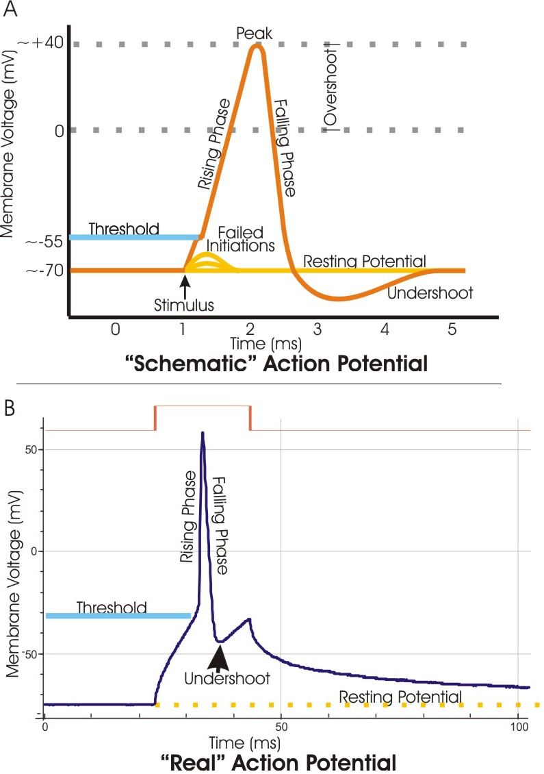
The receptive field is the region of skin or of space where a stimulus changes that neuron’s firing. Fields have shape: a small skin spot, a retinal centre and surround, a cortical bar at one orientation. Transduction makes a graded receptor potential. Dorsal-root and olfactory cells are themselves the afferents, and the potential spreads to a spike trigger. Photoreceptors, hair cells, and taste cells usually do not spike; they change glutamate release onto the next cell.
6.2 Touch and pain #
6.2.1 Mechanoreceptors and the two ascending paths #
Glabrous skin divides low-threshold touch among four endings.
| Receptor | Adaptation | Field | Reports well |
|---|---|---|---|
| Meissner corpuscle | Rapid | Small | Flutter and slip |
| Merkel disc | Slow | Small | Pressure, edges, texture |
| Pacinian corpuscle | Very rapid | Large | High-frequency vibration |
| Ruffini ending | Slow | Large | Skin stretch |
The Pacinian capsule is a mechanical high-pass filter. Strip it, in the classical experiment, and the bare ending adapts much more slowly. The channel did not change. Proprioception comes mainly from muscle spindles and tendon organs (Chapter 7), on fast myelinated axons.
Fine touch, vibration, and proprioception ascend ipsilaterally in the dorsal columns, synapse in the medulla, cross as internal arcuate fibres, and continue in the medial lemniscus to ventral posterior thalamus and postcentral cortex. Pain, temperature, and crude touch synapse in the dorsal horn, cross within a segment or two, and ascend in the spinothalamic tract. A left hemisection, the Brown-Séquard pattern, therefore costs ipsilateral fine touch and proprioception and contralateral pain and temperature below the cut.
6.2.2 Nociceptors and TRP channels #
Nociceptors are free endings of thinly myelinated Aδ axons, for fast sharp pain, and of unmyelinated C axons, for slow burning pain. Many answer to strong mechanical, thermal, and chemical stimuli. TRPV1 opens above about 43 °C and also binds capsaicin and protons, so chilli and damaging heat share a channel on an axon already labelled as pain. TRPM8 opens to cooling below about 25 °C and to menthol.
Injury releases protons, prostaglandins, bradykinin, and nerve growth factor, which lower threshold. A noxious stimulus then hurts more (hyperalgesia), and light touch can hurt (allodynia). Repeated C-fibre input also potentiates dorsal-horn synapses through NMDA receptors, called wind-up while it is brief. Rubbing a knock drives inhibitory interneurons from large touch axons and closes a dorsal-horn gate.
6.3 Vision #
6.3.1 Photoreceptors hyperpolarise #
The retina is central nervous tissue. Rods and cones face the pigment epithelium, which recycles chromophore and absorbs stray light. At the fovea the inner layers are pushed aside and cones are densest: acuity is highest there. The optic disc has no photoreceptors and is the blind spot. Rods can signal single photons and saturate in daylight. Three cone opsins peak near 420, 530, and 560 nm, and colour is a comparison across cones. Rods dominate the periphery, which is why a dim star is easier off the fovea.
Both photoreceptors hyperpolarise to light. In the dark they sit near −40 mV and release glutamate steadily. Light drives them toward about −70 mV and glutamate falls.
6.3.2 The cGMP cascade #
The pigment is an opsin holding 11-cis-retinal. A photon isomerises it to all-trans. In rods, activated rhodopsin turns on transducin, which activates a phosphodiesterase that destroys cGMP. In darkness, cGMP holds cation channels open. Sodium and calcium enter the outer segment — the dark current — while the inner segment pumps sodium out. When cGMP falls, channels close and the cell hyperpolarises.
One rhodopsin activates many transducins, and each enzyme hydrolyses many molecules of cGMP, so a rod reports a single photon. Cones use the same cascade at lower gain and recover faster. Shut-off is active. The pigment epithelium regenerates the chromophore. Rhodopsin kinase and arrestin silence the opsin. Calcium falls as the channels close, guanylyl cyclase speeds up, and gain returns.
6.3.3 On and off pathways, and the path to V1 #
Off bipolar cells express ionotropic glutamate receptors. Glutamate depolarises them in the dark; light, by removing glutamate, hyperpolarises them. They keep the photoreceptor’s sign. On bipolar cells use mGluR6, arranged so that glutamate closes the cation channel TRPM1. They are hyperpolarised in the dark. When light removes glutamate, TRPM1 opens and they depolarise. They invert the sign. On and off ganglion cells therefore report brightening and dimming. Only ganglion cells send spikes over a long distance.
Horizontal cells build centre–surround antagonism. An on-centre ganglion cell is excited by light in a small centre and inhibited by light in the ring. Even illumination of both does little. At the chiasm, nasal axons cross and temporal axons stay. The left visual field falls on the right half of each retina and reaches the right hemisphere. Cutting one optic nerve blinds that eye. Cutting one optic tract, or the cortex it serves, removes the opposite field of both eyes.
The main path for conscious vision is the lateral geniculate nucleus, where the two eyes and the magnocellular and parvocellular streams stay partly apart, then the optic radiation to V1. Other axons reach the superior colliculus, for orienting, and the suprachiasmatic nucleus, for the clock. In V1, Hubel and Wiesel found cells selective for oriented bars, laid out in orientation and ocular-dominance maps.
6.4 Hearing and balance #
6.4.1 Tonotopy and the cochlear amplifier #
The ossicles match the impedance of air to that of cochlear fluid. The eardrum is much larger than the stapes footplate, and the ossicular lever adds a further advantage, so pressure at the oval window is on the order of twenty times the pressure in air. The round window bows out so that incompressible fluid can move. The basilar membrane is narrow and stiff at the base and wider and floppier at the apex. High frequencies peak at the base and low frequencies at the apex, the travelling wave von Békésy described, and that place code is preserved through the cochlear nuclei, the inferior colliculus, and the medial geniculate to primary auditory cortex.
A dead cochlea tunes broadly. About 3,500 inner hair cells supply most of the afferent axons. About 12,000 outer hair cells, in three rows, are the amplifier rather than the main reporters. Prestin changes shape with membrane voltage, so the cell shortens and lengthens and adds energy at the place of the travelling wave.
6.4.2 Tip links, and potassium entry from endolymph #
Stereocilia stand in a height gradient, tied by a tip link of cadherin-23 and protocadherin-15. Bending toward the tallest stereocilium tensions the link and opens a cation channel whose pore subunit is TMC1. The opposite bend closes channels. Calcium that enters resets tip-link tension, in part through myosin, so a steady deflection does not hold the current at its maximum.
The current is potassium, which seems impossible until the compartments are separated. Endolymph, secreted by the stria vascularis, is rich in potassium and is held about +80 mV relative to perilymph. The hair-cell interior is negative to perilymph, about −45 to −55 mV. Across the apical face the inside is therefore well over 100 mV negative to the endolymph, and potassium enters even though it is high on both sides of that membrane. It leaves basolaterally into perilymph.
6.4.3 Canals and otolith organs #
Three semicircular canals on each side sense angular acceleration. At the start of a turn, endolymph lags, the cupula bends, and the bundles deflect. At constant velocity the fluid catches up and the signal fades, so the canals are accelerometers. They work as push–pull pairs: the two horizontal canals, and each anterior canal roughly in the plane of the opposite posterior canal. A turn that excites one member inhibits the other.
The utricle and saccule sense linear acceleration, including gravity. Otoconia load a gel, so a tilt is a sustained stimulus, unlike constant-speed rotation. The utricular macula lies near the horizontal plane and the saccular macula near the vertical.
6.5 Taste and smell #
6.5.1 A short account of both #
Taste, as distinct from flavour, is a small set of qualities: salt, sour, sweet, bitter, and umami. Sweet uses the T1R2–T1R3 pair and umami the T1R1–T1R3 pair. Bitter uses many T2R receptors in one cell, so that cell reports bitter and cannot name the compound. Salt taste is partly an epithelial sodium channel, more clearly so in other mammals than in humans. Sour is a proton taste, with OTOP1 the clearest channel. The cells are epithelial, replaced over days to a couple of weeks, and they synapse on afferents of the facial, glossopharyngeal, and vagus nerves, which end in the nucleus of the solitary tract. Flavour adds retronasal smell and trigeminal irritation.
Each olfactory neuron expresses one receptor gene, from several hundred functional genes in humans and more in many other mammals. That one-receptor rule, from Buck and Axel, is the vertebrate rule of thumb used here. The receptor works through Golf and cAMP, opens a cyclic-nucleotide-gated channel, and lets calcium open a chloride channel. Chloride leaves, because these cells keep internal chloride high, and so adds to the depolarisation. An odorant activates a combination of receptors, and all neurons of one receptor type converge on one glomerulus. Mitral cells then reach piriform cortex without an obligatory thalamic relay, the exception noted in Chapter 5.
6.6 Key terms, summary, and self-check #
Key terms. Labelled line; receptive field; receptor potential; spinothalamic tract; TRPV1; rhodopsin; cGMP; on bipolar; centre–surround; optic chiasm; tonotopy; tip link; endocochlear potential; semicircular canal; otolith; glomerulus.
Summary. Modality is a labelled line, and transduction is a graded receptor potential. Touch receptors split the skin by adaptation and field size; pain crosses in the cord and fine touch in the medulla. Photoreceptors hyperpolarise because light lowers cGMP; on bipolars invert that signal, and ganglion cells report contrast on the way to V1. Frequency is a place on the basilar membrane, tip links open the hair-cell channel, and potassium enters from the positive endolymph. Canals report angular acceleration and otoliths report linear acceleration and tilt. Taste uses a few GPCR and channel qualities. Smell uses one receptor type per neuron and a pattern of glomeruli.
Self-check.
1. Why does a blow to the eye produce a sensation of light?
2. A left spinal hemisection removes pain from which side below the lesion, and fine touch from which side?
3. Light closes a photoreceptor channel. Why does the on bipolar then depolarise?
4. Potassium is high on both sides of the hair cell’s apical membrane. What drives it in?
5. Why can one bitter cell not name the compound, while one glomerulus stands for one receptor type?
—
—
—