
10.1 Conduction and the end-plate #
10.1.1 Multiple sclerosis #
Multiple sclerosis damages central myelin. Peripheral nerves are spared, because their myelin is not the target. Plaques injure oligodendrocytes and their sheaths. The immune effectors are Immunology. A bare stretch of axon leaks current and has a higher capacitance, so local circuit current is spent before the next node. Conduction slows, or it blocks. Warming shortens channel currents and lowers the safety factor further, which is why a rise in temperature can uncover symptoms, the effect associated with Uhthoff.
Conduction can return if the stretch is remyelinated, often with a thinner and shorter sheath, or if sodium channels spread along the bare membrane and support continuous conduction. Axons are also cut in plaques, and accumulated axonal loss, not the myelin gap alone, accounts for much of the lasting deficit.
10.1.2 Myasthenia gravis #
Myasthenia gravis is a disease of the end-plate, not of the motor neuron and not of the filament. In most people with it, antibodies bind the muscle nicotinic receptor. They cross-link receptors and speed their destruction, they fix complement so that the folds are damaged, and they can block the acetylcholine site. Each quantum opens fewer channels. The end-plate potential, normally far above threshold, shrinks toward it.
Early in a contraction the fibre may still fire. With repeated use, release depresses slightly, as it does at a healthy junction. There the safety factor absorbs the fall. Here it does not, successive end-plate potentials miss threshold, and the muscle fatigues. A rest lets release recover. A smaller group of patients have antibodies to MuSK instead, so receptors may be present and still fail to cluster under the nerve, the developmental step of Chapter 8 left unfinished. Lambert–Eaton myasthenic syndrome is the presynaptic contrast: antibodies to P/Q-type calcium channels reduce quantal content, and a brief effort can facilitate release because residual calcium builds.
10.2 Selection, and excitation against inhibition #
10.2.1 Parkinson disease #
The motor picture discussed here follows loss of dopamine neurons in the substantia nigra pars compacta, and so of their supply to the dorsal striatum. Remaining cells often contain Lewy bodies rich in α-synuclein. Why those neurons die is not settled. Mitochondrial stress, failed protein clearance, and spread of misfolded α-synuclein are all part of the argument.
Without dopamine the direct pathway responds less to cortex and the indirect pathway is less suppressed. The internal pallidum therefore fires too much, the thalamus is over-inhibited, and cortex is under-driven. Movement is hard to start and, once started, small and slow. The rigidity is not the velocity-dependent spasticity of a corticospinal lesion. A resting tremor is typical. The pathway diagram accounts for the poverty of movement more directly than for that oscillation.
10.2.2 Epilepsy #
A seizure is a transient, excessive, synchronised discharge. Epilepsy is a tendency to further unprovoked seizures. It is not one channel and not one disease. The common theme is a failure of the balance between excitation and inhibition, or of the brakes that should end a burst. Loss of SCN1A in inhibitory interneurons, as in Dravet syndrome, causes seizures by weakening that brake.
In a focal cortical seizure, cells undergo a paroxysmal depolarising shift: a long depolarisation, fed by recurrent excitation, with a burst of spikes on top. Potassium currents and nearby inhibitory cells normally cut it short. If GABA is weak, or if NMDA-dependent potentiation has strengthened the recurrent synapses, the burst spreads across a map and can appear as a march of movement from one body part to the next.
Absence seizures use a different circuit. Thalamic relay and reticular cells rebound through T-type calcium channels and lock the cortex into a rhythmic discharge, with a brief lapse of awareness. This is spindle machinery at the wrong gain, not a cortical paroxysmal shift.
10.3 Alzheimer disease #
10.3.1 Plaques, tangles, and an argued chain #
In its typical course, Alzheimer disease begins as a failure of episodic memory and later widens. Entorhinal cortex and hippocampus are involved early, which fits Chapter 9, and association cortex follows. The classical lesions are extracellular plaques of amyloid-β and intracellular neurofibrillary tangles. Hyperphosphorylated tau leaves microtubules and assembles into the paired helical filaments of those tangles. Loss of synapses tracks the cognitive decline more closely than a simple count of plaques does.
Amyloid-β is cut from its precursor by β-secretase and by γ-secretase, whose catalytic subunit is presenilin. A cut by α-secretase falls inside the peptide and prevents it. Mutations in the precursor or in presenilin cause rare early-onset dominant disease and increase the aggregation-prone peptide.
The amyloid cascade hypothesis says that the peptide causes the rest, and that removing it should stop the disease. Familial genetics support an upstream place for it in those families. The chain is still argued for the common sporadic disease. Plaques occur in some older people whose cognition holds. Tau pathology and synapse loss follow the decline more closely than plaque burden does. Vascular injury, inflammation, and loss of basal-forebrain cholinergic cells all belong in the picture, and none has been shown to be the single start of sporadic disease.
10.4 Key terms, summary, and self-check #
Key terms. Conduction block; safety factor; myasthenia gravis; MuSK; Lambert–Eaton; substantia nigra; Lewy body; paroxysmal depolarising shift; amyloid-β; neurofibrillary tangle.
Summary. Multiple sclerosis removes central myelin, so saltatory conduction fails and axons are later lost. Myasthenia gravis removes or mis-clusters nicotinic receptors, so the end-plate potential falls below threshold with use; Lambert–Eaton syndrome instead cuts calcium-dependent release. Parkinson disease removes nigral dopamine, the internal pallidum over-inhibits the thalamus, and movement is not selected. Epilepsy collects failures of excitation–inhibition balance, from cortical bursts to thalamocortical absence rhythms. Alzheimer disease degenerates declarative memory circuitry and accumulates amyloid-β and tau; familial genetics implicate amyloid-β, and the sporadic causal chain remains argued.
Self-check.
1. Why can a demyelinated axon fail even though sodium channels have not all been removed?
2. Why does myasthenic weakness grow during a sustained effort, rather than being fixed from the first twitch?
3. Which step of transmission do the antibodies remove in Lambert–Eaton syndrome, as against typical myasthenia?
4. Why does loss of nigral dopamine increase, rather than decrease, inhibitory output from the internal pallidum?
5. What do familial presenilin mutations imply about amyloid-β, and why is that not a finished cause for sporadic disease?
—
# Closing: how the chapters fit together
A neuron is a cell with a long axon and a threshold. Chapter 1 gives that cell, the glia that stabilise its surroundings, and the myelin and endothelial barrier of a vertebrate brain. Chapter 2 turns a potassium resting potential into a sodium–potassium spike that normally runs one way, and faster if the axon is thick or myelinated. Chapter 3 turns that spike into quantal secretion, judged at the initial segment.
Chapter 4 names the receptors that decide excitation, inhibition, or a change of gain. The NMDA receptor is the coincidence detector that Chapter 9 spends. Chapter 5 sets the junctions into a reflex, into regions defined by a computation rather than a slogan, into two autonomic outflows, and into a fluid space around the whole. Chapters 6 and 7 are labelled, mapped axons used to sense and to move: a receptor potential in skin, retina, and hair cell, and a command that has passed spinal pattern, brainstem posture, cortex, cerebellar correction, and basal-ganglia selection.
Chapters 8 and 9 build the wiring and let activity store a change. Chapter 10 is myelin, the end-plate, nigral dopamine, inhibition, and Alzheimer proteins failing. Revision runs in that order. Without the initial segment, the direction of the sodium current, and what the NMDA receptor is waiting for, a map or a lesion or a memory experiment cannot yet be read.
Revision checklist #
1. Draw a neuron, mark the initial segment, and name the motor and the microtubule polarity of anterograde transport.
2. From the Nernst potentials of sodium and potassium, say why rest lies near the potassium potential and why the spike moves toward the sodium potential and then away.
3. Use the refractory periods to explain one-way travel, and say how myelin changes speed without changing the ions.
4. Order a chemical synapse from the presynaptic spike to termination, and say what a miniature end-plate potential is.
5. Separate curare, botulinum toxin, and acetylcholinesterase block by the single step each one removes.
6. Explain the magnesium block, and why a weak input paired with depolarisation potentiates at CA1.
7. For heart rate and for the pupil, name the transmitter and the receptor class used by each autonomic division.
8. Explain why a photoreceptor hyperpolarises in light, and how an on bipolar inverts that signal. Separately, explain why potassium enters a hair cell from endolymph.
9. In one line each, contrast an upper motor neuron lesion, a lower motor neuron lesion, a cerebellar lesion, and the poverty of movement after dopamine loss.
10. State what H.M. could still learn, what he could not newly form, and why that separates the site of consolidation from the permanent store.
Selected sources #
These notes are an original teaching summary, not a transcript of any textbook.
- Kandel ER, Koester JD, Mack SH, Siegelbaum SA (eds). Principles of Neural Science. 6th edition. McGraw Hill, 2021. Earlier editions, edited with Schwartz and Jessell, are the same tradition.
- Purves D, Augustine GJ, Fitzpatrick D, Hall WC, LaMantia A-S, White LE (eds). Neuroscience. Oxford University Press / Sinauer. Recent editions.
- Nicholls JG, Martin AR, Wallace BG, Fuchs PA. From Neuron to Brain. 5th edition. Sinauer Associates, 2012.
- Hodgkin AL, Huxley AF. A quantitative description of membrane current and its application to conduction and excitation in nerve. Journal of Physiology 117:500–544 (1952).
- Bliss TVP, Lømo T. Long-lasting potentiation of synaptic transmission in the dentate area of the anaesthetized rabbit following stimulation of the perforant path. Journal of Physiology 232:331–356 (1973).
—
—
# Part C. Visual Figures & Scientific Diagrams Gallery
Comprehensive high-resolution visual reference repository for Neurobiology from the B10logy Visual Atlas.
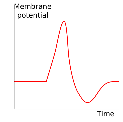
Action Potential Basic Shape
—
# Part C. Visual Figures & Scientific Diagrams Gallery
Essential high-resolution reference diagrams for Neurobiology from the B10logy Visual Atlas.
