
9.1 Chemical Analysis: Acid-Soluble Pool (Biomicromolecules) vs Acid-Insoluble Pellets (Biomacromolecules) #
To find out what kinds of organic compounds are present in living organisms, biologists perform a simple chemical analysis on living tissues, such as a piece of vegetable (like a carrot or potato) or a small piece of animal tissue (such as liver).
The tissue is ground thoroughly in trichloroacetic acid (Cl3CCOOH) using a mortar and pestle. This produces a thick slurry. When this slurry is filtered through a cheesecloth or cotton mesh, it separates into two distinct fractions:
- The Filtrate (Acid-Soluble Pool): This is the liquid that passes freely through the filter cloth. It roughly represents the composition of the cytoplasm. It contains thousands of small organic molecules called biomicromolecules with molecular weights ranging strictly between 18 and 800 Daltons (Da), such as amino acids, simple sugars (monosaccharides), and nucleotides.
- The Retentate (Acid-Insoluble Fraction): This is the pellet that remains trapped on the filter cloth. It represents macromolecules derived from the cytoplasm and cellular organelles. It contains four major classes of biomacromolecules—proteins, nucleic acids, polysaccharides, and lipids—all of which (except lipids) have large molecular weights of 10,000 Daltons and above.
9.2 Primary vs Secondary Metabolites (Alkaloids, Flavonoids, Terpenoids, Toxins, Lectins) #
When living tissues are analyzed, thousands of organic compounds are discovered. In animal tissues, all the compounds noticed—such as sugars, amino acids, fatty acids, glycerol, and nucleotides—are called primary metabolites because they have identifiable functions and play known, direct roles in normal physiological and metabolic processes.
However, when we analyze plant, fungal, and microbial cells, we encounter thousands of additional compounds such as alkaloids, flavonoids, rubber, essential oils, antibiotics, colored pigments, scents, gums, and spices. These are called secondary metabolites. While many of them do not have obvious, direct roles in the host plant’s primary growth or development, many are ecologically vital (defending against herbivores) and commercially valuable for human welfare.
| Category of Secondary Metabolite | NCERT Representative Examples | Biological / Human Significance |
|---|---|---|
| Pigments | Carotenoids, Anthocyanins | Light absorption, floral coloration for pollinators |
| Alkaloids | Morphine, Codeine | Potent analgesics derived from opium poppy |
| Terpenoids | Monoterpenes, Diterpenes | Aromatic defense compounds and resin components |
| Essential Oils | Lemon grass oil | Fragrances, anti-microbial plant defenses |
| Toxins | Abrin (from Abrus), Ricin (from Castor) | Ribosome-inactivating defensive poisons |
| Lectins | Concanavalin A | Carbohydrate-binding agglutination proteins |
| Drugs | Vinblastin (anticancer), Curcumin (anti-inflammatory) | Modern therapeutics from Catharanthus & turmeric |
| Polymeric Substances | Rubber, Gums, Cellulose | Structural sealants, commercial elastomers |
9.3 Proteins: Amino Acid Building Blocks, Peptide Bonds & 1°, 2°, 3°, 4° Structural Levels #
Biomacromolecules are large polymeric compounds formed by the covalent polymerization of monomeric building blocks. Polysaccharides are long chains of monosaccharide sugars linked by glycosidic bonds:
- Cellulose: A straight, unbranched homopolymer made entirely of glucose monomers linked by β-1,4-glycosidic bonds. Plant cell walls are made of cellulose. Cotton fiber is almost pure cellulose. Paper made from plant pulp is cellulose. Notice: Cellulose does not contain complex helices and cannot hold iodine molecules, so it does not give a blue color with iodine!
- Starch: A polymeric storehouse of energy in plant tissues. It consists of two components: unbranched amylose (helical) and branched amylopectin. Starch forms secondary helical structures that can hold iodine (I2) molecules in its helical interior, producing an intense deep blue color.
- Glycogen: The reserve carbohydrate storage polysaccharide in animals and fungi (‘animal starch’). It is highly branched, resembling amylopectin. In a glycogen chain, the right end is called the reducing end and the left end is the non-reducing end.
- Inulin: A homopolymer of fructose used clinically to assess glomerular filtration rate (GFR) in kidneys.
- Complex Exoskeletal Polysaccharides: Chitin is a complex homopolymer of amino sugars, specifically N-acetyl glucosamine (NAG), forming the tough exoskeletons of arthropods and cell walls of fungi.
9.4 Polysaccharides: Starch, Glycogen, Cellulose & Glycosidic Bonds #
Proteins are polypeptide chains composed of amino acids linked together by peptide bonds. Since there are 20 different amino acids occurring in proteins, a protein is never a homopolymer; it is always a heteropolymer of amino acids. Dietary proteins are the source of essential amino acids that cannot be synthesized by the human body and must be supplied through food.
Proteins exhibit four progressive levels of structural organization:
- Primary Structure: The exact linear sequence of amino acids in a polypeptide chain. The first amino acid at the left end has a free amino group and is called the N-terminal amino acid; the last amino acid at the right end has a free carboxyl group and is called the C-terminal amino acid.
- Secondary Structure: The folding of the linear polypeptide chain into localized regular conformations via hydrogen bonding between peptide backbone groups: the α-helix (strictly right-handed helices in proteins) and the β-pleated sheet.
- Tertiary Structure: The long protein chain folds extensively upon itself like a hollow woolen ball, creating a compact three-dimensional globular architecture. Tertiary structure is stabilized by hydrophobic interactions, ionic bonds, hydrogen bonds, and covalent disulfide (-S-S-) bridges. Tertiary structure is absolutely necessary for the biological activities of proteins, creating specific clefts called active sites for enzymes.
- Quaternary Structure: Seen in proteins composed of more than one polypeptide subunit. The spatial assembly of individual folded subunits forms quaternary structure. For example, adult human hemoglobin (Hb) consists of 4 subunits: two identical α-chains and two identical β-chains (α2β2).
- Collagen is the most abundant protein in the entire animal world.
- RuBisCO (Ribulose bisphosphate carboxylase-oxygenase) is the most abundant protein in the whole of the biosphere!
9.5 Lipids: Fatty Acids, Glycerol, Phospholipids (Lecithin) & Sterols #
Nucleic acids (DNA and RNA) are macromolecules found in the acid-insoluble fraction of all living cells. They are polynucleotides composed of repeating monomeric units called nucleotides. A nucleotide consists of three chemically distinct components:
- A heterocyclic nitrogenous base: Classified into double-ringed Purines (Adenine [A] and Guanine [G]) and single-ringed Pyrimidines (Cytosine [C], Uracil [U] in RNA, and Thymine [T] in DNA).
- A pentose monosaccharide sugar: Ribose in ribonucleic acid (RNA) or 2′-deoxyribose in deoxyribonucleic acid (DNA). When a nitrogenous base is attached to a pentose sugar via an N-glycosidic bond, it is called a nucleoside (e.g., Adenosine, Guanosine, Cytidine, Uridine, Thymidine).
- A phosphate group: When a phosphate group is esterified to the 5′-OH of a nucleoside, it forms a complete nucleotide (e.g., Adenylic acid, Guanylic acid, Uridylic acid, Cytidylic acid, Thymidylic acid).
In 1953, James Watson and Francis Crick proposed the classic double-helix model of B-DNA:
- DNA consists of two polynucleotide strands running in antiparallel directions (one 5’→3′, the other 3’→5′).
- The sugar-phosphate backbone forms the outer structural rail, while nitrogenous bases project inward perpendicular to the axis.
- Bases pair specifically via hydrogen bonds: Adenine pairs with Thymine with two hydrogen bonds (A = T), and Guanine pairs with Cytosine with three hydrogen bonds (G ≡ C).
- The pitch of the B-DNA helix is 3.4 nm (34 Å) with roughly 10 base pairs per turn, meaning the distance between adjacent base pairs is 0.34 nm (3.4 Å). The plane of one base pair stacks over the other in the double helix, conferring thermodynamic stability.
9.6 Nucleic Acids: Nucleosides, Nucleotides, Phosphodiester Bonds & Double Helix Watson-Crick B-DNA #
Biopolymers are assembled by covalent linkages forged through condensation (dehydration) reactions between adjacent monomer units:
- Peptide Bond: Links amino acids in a protein. Formed when the carboxyl group (-COOH) of one amino acid reacts with the amino group (-NH2) of the next amino acid, eliminating a molecule of water (-CO-NH-).
- Glycosidic Bond: Links individual monosaccharides in a polysaccharide. Formed between two carbon atoms of two adjacent monosaccharide rings with the loss of water (e.g., α-1,4-glycosidic bond in maltose and starch).
- Phosphodiester Bond: Links nucleotides in a nucleic acid strand. The phosphate group connects the 3′-carbon of one sugar of one nucleotide to the 5′-carbon of the sugar of the succeeding nucleotide. Because there is an ester bond on each side of the phosphate group, it is called a phosphodiester bond.
- Ester Bond in Lipids: In triglycerides, three fatty acid molecules are esterified to the three hydroxyl groups of one glycerol (trihydroxy propane) molecule via ester bonds.
9.7 Enzymology: Activation Energy, Active Site, Factors Affecting Activity (pH, Temp, Substrate Conc) #
In every living cell, thousands of biomolecules are present in specific metabolic concentrations. These molecules are never static; they are constantly being broken down and transformed into some other biomolecules, and also made from other biomolecules. This continuous flow and chemical transformation is called turnover, and all these chemical reactions together constitute metabolism.
Metabolic transformations never occur in isolation; they are always linked to other reactions in orderly series called metabolic pathways (similar to city automobile traffic). Metabolic pathways can be:
- Anabolic (Biosynthetic) Pathways: Simpler structures combine to form more complex molecules, consuming energy (e.g., synthesis of proteins from amino acids; requires ATP).
- Catabolic (Degradative) Pathways: Complex molecules are dismantled into simpler fragments, releasing energy (e.g., glycolysis breaking glucose into lactic acid or pyruvate; yields ATP). The universal energy currency of cellular work is Adenosine Triphosphate (ATP).
A system at thermodynamic equilibrium cannot perform work. Living organisms work continuously to maintain homeostasis; therefore, living systems cannot afford to reach equilibrium. The living state is a non-equilibrium steady state capable of performing work. Metabolism provides the continuous energy input required to prevent the living system from falling into thermodynamic equilibrium (death).
9.8 Enzyme Inhibition: Competitive (Km increases, Vmax constant e.g. Malonate vs Succinate Dehydrogenase) vs Non-competitive #
Almost all enzymes are proteins (with rare catalytic RNA exceptions called ribozymes). Like all proteins, an enzyme has primary, secondary, and tertiary structures. The folded tertiary structure forms specific physical crevices or pockets called active sites into which the substrate fits, catalyzing chemical transformations at extraordinary velocities.
An enzyme dramatically accelerates a reaction by lowering the activation energy barrier required to transition the substrate into high-energy transition state complexes:
- Factors Affecting Enzymatic Activity:
- Temperature & pH: Enzymes display maximum activity at optimum temperature and optimum pH. Activity declines above and below the optimum; low temperatures temporarily inactivate enzymes, while high temperatures permanently destroy enzymatic activity by heat-induced protein denaturation.
- Substrate Concentration [S]: As [S] increases, reaction velocity (V) rises rapidly until it reaches a maximum velocity (Vmax). Once all enzyme active sites are saturated, further increase in [S] cannot increase velocity. The Michaelis-Menten constant (Km) is the substrate concentration at which the reaction velocity is half of Vmax (Vmax/2). Lower Km indicates higher enzyme-substrate affinity.
- Enzyme Inhibition: When the activity of an enzyme is shut off by a specific chemical, the process is called inhibition. In competitive inhibition, the inhibitor closely resembles the substrate in molecular structure and competes for the active site. For example, malonate closely resembles succinate and competitively inhibits succinate dehydrogenase. Competitive inhibition increases apparent Km while Vmax remains unchanged!
- Enzyme Classification (6 Classes of Enzymes):
- Oxidoreductases / Dehydrogenases: Catalyze oxidation-reduction between two substrates.
- Transferases: Catalyze transfer of a group (other than hydrogen) between substrates.
- Hydrolases: Catalyze hydrolysis of ester, ether, peptide, glycosidic, or C-C bonds.
- Lyases: Catalyze removal of groups from substrates by mechanisms other than hydrolysis, leaving double bonds.
- Isomerases: Catalyze interconversion of optical, geometric, or positional isomers.
- Ligases: Catalyze the linking together of two compounds (e.g., C-O, C-S, C-N, P-O bonds) using ATP.
- Cofactors: Non-protein chemical components bound to enzymes to make them catalytically active. The catalytic protein part alone is called the apoenzyme. Cofactors include:
- Prosthetic Groups: Tightly and permanently bound organic compounds (e.g., Heme in peroxidase and catalase).
- Co-enzymes: Transiently bound organic compounds, often vitamins (e.g., NAD and NADP contain the vitamin niacin).
- Metal Ions: Form coordination bonds with active site side chains (e.g., Zinc (Zn2+) is a cofactor for the proteolytic enzyme carboxypeptidase and carbonic anhydrase).
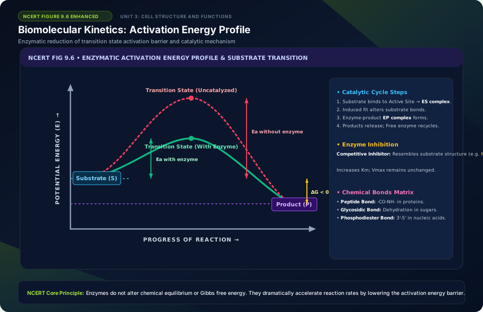
Biomolecular Kinetics: Activation Energy Profile
🚨 Examiner Traps & Common Misconceptions
✅ Reality: Lipids have low molecular weights (<800 Da), but appear in the acid-insoluble fraction because they form insoluble vesicles and membrane fragments during tissue homogenization.
✅ Reality: Competitive inhibitors do NOT lower Vmax because adding enough substrate outcompetes the inhibitor! They only increase Km.
💡 Memory Hack & Mnemonic
Secondary Metabolites Toxins vs Lectins: ‘Ricin and Abrin Attack (Toxins)’; ‘Concanavalin A Loves sugars (Lectin)’.
⭐ 10/10 Active Recall Self-Check: Chapter 9
Test your mastery before closing this lecture note. Can you answer these without looking back?
- What are the primary diagnostic characteristics that define Biomolecules in the NCERT syllabus?
- How do the physiological or anatomical structures covered in this chapter adapt the organism to its environment?
- What are the most common trick questions examiners create based on the exceptions in this chapter?
- Can you state the core mnemonic formula for remembering the sequential stages or components described above?
⭐ 10/10 Active Recall Self-Check: Chapter 9
Test your mastery before closing this lecture note. Can you answer these without looking back?
- What are the primary diagnostic characteristics that define Biomolecules in the NCERT syllabus?
- How do the physiological or anatomical structures covered in this chapter adapt the organism to its environment?
- What are the most common trick questions examiners create based on the exceptions in this chapter?
- Can you state the core mnemonic formula for remembering the sequential stages or components described above?