
8.1 Cell Theory: Schleiden, Schwann & Rudolf Virchow (‘Omnis cellula-e cellula’) #
When you examine any living organism under a microscope, you discover that whether it is a giant sequoia tree or an invisible bacterium, life is packaged into microscopic compartments called cells. A cell is the fundamental structural and functional unit of all living organisms because anything less than a complete cell does not ensure independent living.
Anton von Leeuwenhoek first saw and described a live cell under his handcrafted microscope. Later, Robert Brown discovered the nucleus. The formulation of the Cell Theory was a collaborative historical milestone:
- In 1838, Matthias Schleiden, a German botanist, examined a large number of plants and concluded that all plants are composed of different kinds of cells that form the tissues of the plant.
- In 1839, Theodore Schwann, a British zoologist, studied animal cells and reported that cells had a thin outer layer (now known as the plasma membrane). Studying plant tissues, he also discovered that the presence of a cell wall is a unique character of plant cells. Schwann proposed the hypothesis that the bodies of animals and plants are composed of cells and products of cells.
- However, Schleiden and Schwann did not explain how new cells were formed. In 1855, Rudolf Virchow first explained that cells divide and new cells are formed from pre-existing cells: Omnis cellula-e cellula.
- All living organisms are composed of cells and products of cells.
- All cells arise from pre-existing cells (Omnis cellula-e cellula).
8.2 Prokaryotic Cell Architecture: Cell Envelope, Mesosomes, Plasmids & 70S Ribosomes #
A typical cell possesses an outer boundary, semi-fluid matrix called cytoplasm where vital cellular activities and chemical reactions occur, and genetic material. Based on the presence or absence of a membrane-bound nucleus and specialized organelles, cells are classified into two major categories:
- Prokaryotic Cells: Cells that lack a membrane-bound nucleus and membrane-bound organelles. Their genetic material lies naked in the cytoplasm as a nucleoid (represented by Bacteria, Blue-green algae, Mycoplasma, and PPLO).
- Eukaryotic Cells: Cells possessing a true membrane-bound nucleus with chromosomes, and extensive internal compartmentalization by membrane-bound organelles (Endoplasmic reticulum, Golgi apparatus, lysosomes, mitochondria, and plastids). Found in Protists, Fungi, Plants, and Animals.
Cells differ dramatically in size, shape, and activities. The smallest known cells are Mycoplasma (only 0.3 µm in length), whereas typical bacteria measure 3 to 5 µm. The largest isolated single cell is the egg of an ostrich. Among human cells, red blood cells (RBCs) are round and biconcave with a diameter of ~7.0 µm, while nerve cells (neurons) are some of the longest cells, reaching up to 1 meter in length.
8.3 Eukaryotic Plasma Membrane: Fluid Mosaic Model of Singer & Nicolson (1972) #
Prokaryotic cells are structurally simpler than eukaryotic cells, but metabolically remarkably diverse. Most prokaryotes, particularly bacteria, have a tightly bound three-layered cell envelope:
- Glycocalyx: Outermost coating of macromolecules. It can be a loose, non-rigid gelatinous sheath called a slime layer, or a thick, tough, rigid protective layer called a capsule (which prevents desiccation and evades host immune phagocytosis).
- Cell Wall: Middle rigid layer composed of peptidoglycan (murein) that determines cell shape and prevents the bacterium from bursting or collapsing under osmotic pressure. Bacteria are classified into Gram-positive and Gram-negative based on cell envelope differences and their response to Gram staining.
- Plasma Membrane: Innermost selectively permeable lipid bilayer that interacts with the outside environment.
A special membranous structure characteristic of prokaryotes is the mesosome, formed by invaginations of the plasma membrane into the cell in the form of vesicles, tubules, and lamellae. Mesosomes help in cell wall synthesis, DNA replication and distribution to daughter cells, respiration, and increasing surface area of the enzymatic plasma membrane.
Motile bacteria possess flagella composed of flagellin protein with three parts: filament, hook, and basal body. Surface appendages also include pili (elongated tubular structures made of pilin protein involved in conjugation) and fimbriae (small bristle-like fibers helping bacteria attach to rocks in streams or host tissues). Prokaryotic ribosomes are 70S (composed of 50S large and 30S small subunits).
8.4 The Endomembrane System: Endoplasmic Reticulum (RER & SER), Golgi Apparatus, Lysosomes & Vacuoles #
The detailed structure of the eukaryotic plasma membrane was revealed only after the advent of the electron microscope in the 1950s. Chemical studies on human red blood cell membranes showed that the membrane is composed predominantly of lipids (40%) and proteins (52%), along with carbohydrates (8%).
The lipids are arranged as a phospholipid bilayer. The polar, hydrophilic head of each phospholipid molecule faces outward toward the aqueous environment, while the nonpolar, hydrophobic hydrocarbon tails of fatty acids face inward toward the center, shielded from aqueous contact. In addition to phospholipids, membranes contain cholesterol, which regulates membrane fluidity and stability.
In 1972, Singer and Nicolson proposed the universally accepted Fluid Mosaic Model of the plasma membrane. According to this model, the quasi-fluid nature of the lipid bilayer enables the lateral movement of proteins within the overall bilayer. This ability to move within the membrane is measured as its fluidity. Membrane proteins are categorized into:
- Integral (Intrinsic) Proteins: Partially or totally buried deep within the lipid bilayer (e.g., transmembrane channel proteins and carrier pumps).
- Peripheral (Extrinsic) Proteins: Lie loosely on the outer or inner surface of the membrane and are easily extracted.
The plasma membrane is selectively permeable. Transport occurs via:
- Passive Transport: Movement of neutral solutes along the concentration gradient (from higher to lower concentration) without any expenditure of energy (ATP). Includes simple diffusion and osmosis (water movement across a semi-permeable membrane). Polar molecules require membrane carrier proteins for facilitated diffusion without ATP.
- Active Transport: Movement of ions or molecules across the membrane against their concentration gradient (from lower to higher concentration). This uphill transport strictly requires metabolic energy in the form of ATP (e.g., the Na+/K+ ATPase pump, which pumps 3 Na+ out and 2 K+ in per ATP hydrolyzed).
8.5 Semiautonomous Energy Organelles: Mitochondria (Cristae, ATP Synthase) & Plastids (Chloroplasts, Thylakoids) #
A non-living rigid structure called the cell wall forms an outer covering for the plasma membrane of fungi and plants. In algae, the cell wall is composed of cellulose, galactans, mannans, and calcium carbonate, whereas in higher plants it consists of cellulose, hemicellulose, pectins, and proteins. Neighboring plant cells are cemented together by the middle lamella (composed of calcium and magnesium pectate) and traversed by cytoplasmic channels called plasmodesmata.
Inside the eukaryotic cell, while many organelles have distinct functions, several are grouped together into the Endomembrane System because their functions are intimately coordinated: Endoplasmic Reticulum (ER), Golgi Apparatus, Lysosomes, and Vacuoles (Mitochondria, chloroplasts, and peroxisomes are not part of the endomembrane system because their functions are uncoordinated with these):
- Endoplasmic Reticulum (ER): Network of tiny tubular structures scattered in the cytoplasm:
- Rough Endoplasmic Reticulum (RER): Bears 80S ribosomes on its outer surface. Actively involved in protein synthesis and secretion; continuous with the outer nuclear membrane.
- Smooth Endoplasmic Reticulum (SER): Lacks ribosomes. Major site for synthesis of lipids, phospholipids, and steroid hormones (in animal cells), as well as calcium sequestration and detoxification.
- Golgi Apparatus: Discovered by Camillo Golgi (1898) as densely stained reticular structures near the nucleus. Consists of flat, disc-shaped sacs or cisternae (0.5 to 1.0 µm diameter) arranged concentrically. Has a convex, forming cis-face (receives vesicles from ER) and a concave, maturing trans-face (releases modified secretory vesicles). Primary function: Packaging, modification, and sorting of proteins and lipids into glycoproteins and glycolipids (glycosidation and glycosylation).
- Lysosomes: Membrane-bound vesicular structures formed by the packaging process in the Golgi apparatus. Rich in all types of hydrolytic enzymes (hydrolases—lipases, proteases, carbohydrases, nucleases) optimally active at an acidic pH (~5.0) maintained by proton pumps. Capable of digesting foreign pathogens and damaged organelles (‘suicide bags’).
- Vacuoles: Membrane-bound space in cytoplasm bound by a single semi-permeable membrane called the tonoplast. In mature plant cells, vacuoles can occupy up to 90% of cellular volume. The tonoplast actively pumps ions into the vacuole against concentration gradients. In Amoeba, a contractile vacuole is vital for osmoregulation and excretion, while in protists, food vacuoles engulf nutrients.
8.6 Ribosomes (80S Eukaryotic), Cytoskeleton (Microtubules, Microfilaments) & Cilia/Flagella (9+2 Axoneme) #
Mitochondria and Chloroplasts are specialized energy-transducing eukaryotic organelles possessing their own circular double-stranded DNA, 70S ribosomes, and RNA components, allowing them to synthesize some of their own proteins. Hence, they are called semi-autonomous organelles and divide by fission.
- Mitochondria (The ‘Powerhouses of the Cell’): Double membrane-bound organelle. The outer membrane is smooth, while the inner membrane forms numerous complex infoldings called cristae that increase surface area for oxidative phosphorylation. The inner compartment is filled with a dense homogeneous gel called the matrix containing Krebs cycle enzymes, a single circular dsDNA molecule, and 70S ribosomes. Studded on the cristae are F0-F1 ATP synthase complexes (oxysomes) that generate cellular ATP via aerobic respiration.
- Plastids: Found in all plant cells and euglenoids. Categorized into:
- Leucoplasts: Colorless plastids storing nutrients: Amyloplasts (store starch, e.g., potato), Elaioplasts (store oils and fats), and Aleuroplasts (store proteins).
- Chromoplasts: Contain fat-soluble carotenoid pigments (carotene, xanthophylls) giving flowers and fruits yellow, orange, or red colors.
- Chloroplasts: Double membrane-bound organelles containing Chlorophyll and carotenoids that trap solar energy for photosynthesis. The space limited by the inner membrane is called the stroma (containing dark-reaction Calvin cycle enzymes, circular DNA, and 70S ribosomes). Organized inside the stroma are flattened membranous sacs called thylakoids arranged in stacks like piles of coins called grana, interconnected by stroma lamellae.
8.7 The Nucleus: Nuclear Envelope, Chromatin, Nucleolus & Chromosome Morphology (Centromere, Kinetochore) #
The cytoplasm of eukaryotic cells is structurally reinforced by an elaborate internal network of filamentous proteinaceous structures called the cytoskeleton. The cytoskeleton consists of three major elements: microtubules (composed of tubulin protein), microfilaments (composed of actin), and intermediate filaments. Functions: mechanical support, motility, maintenance of cell shape, and intracellular transport.
- Cilia and Flagella: Hair-like outgrowths of the cell membrane. Cilia are small, working like oars causing movement of the cell or surrounding fluid; flagella are longer and responsible for cell locomotion. Both are covered by plasma membrane. Their core, called the axoneme, possesses 9 doublets of radially arranged peripheral microtubules and a pair of centrally located microtubules: the classic (9 + 2) axonemal arrangement. The central tubules are enclosed by a central sheath connected to peripheral doublets by radial spokes. Cilia and flagella emerge from centriole-like basal bodies.
- Centrosome and Centrioles: An organelle usually containing two cylindrical structures called centrioles that lie perpendicular to each other, surrounded by amorphous pericentriolar materials. Each centriole is composed of 9 evenly spaced peripheral triplets of tubulin protein with no central microtubule, forming a (9 + 0) cartwheel pattern. Centrioles form the basal body of cilia/flagella and organize the spindle apparatus that guides chromosome separation during animal cell division.
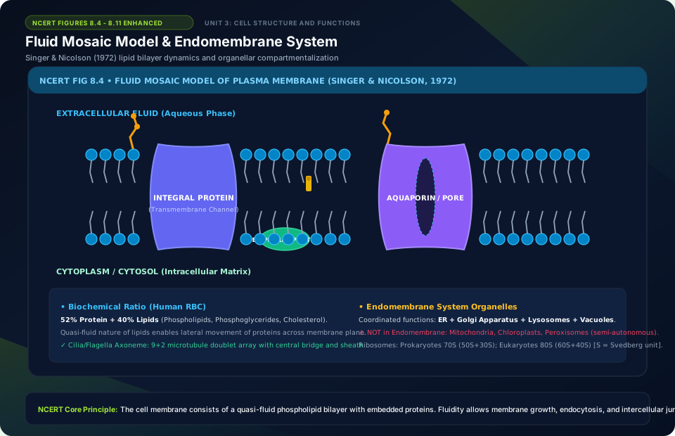
Fluid Mosaic Model & Endomembrane System
🚨 Examiner Traps & Common Misconceptions
✅ Reality: They are semiautonomous endosymbionts: they possess their own circular DNA and 70S ribosomes, but depend on nuclear genes for most of their structural proteins and enzymes!
✅ Reality: Prokaryotes have zero membrane-bound organelles. Respiration occurs at plasma membrane folds called mesosomes.
💡 Memory Hack & Mnemonic
Endomembrane System Organelles: ‘Every Girl Loves Vacation’ (ER, Golgi, Lysosome, Vacuole. Mitochondria, Chloroplasts, and Peroxisomes are NOT part of it!).
⭐ 10/10 Active Recall Self-Check: Chapter 8
Test your mastery before closing this lecture note. Can you answer these without looking back?
- What are the primary diagnostic characteristics that define Cell: The Unit of Life in the NCERT syllabus?
- How do the physiological or anatomical structures covered in this chapter adapt the organism to its environment?
- What are the most common trick questions examiners create based on the exceptions in this chapter?
- Can you state the core mnemonic formula for remembering the sequential stages or components described above?
⭐ 10/10 Active Recall Self-Check: Chapter 8
Test your mastery before closing this lecture note. Can you answer these without looking back?
- What are the primary diagnostic characteristics that define Cell: The Unit of Life in the NCERT syllabus?
- How do the physiological or anatomical structures covered in this chapter adapt the organism to its environment?
- What are the most common trick questions examiners create based on the exceptions in this chapter?
- Can you state the core mnemonic formula for remembering the sequential stages or components described above?