
7.1 Epithelial Tissue: Simple (Squamous, Cuboidal, Columnar, Ciliated) & Compound Epithelium #
In multicellular animals, a group of similar cells along with intercellular substances perform a specific function. Such an organization is called a tissue. Animal tissues are categorized into four fundamental types based on structure and embryonic origin:
- Epithelial Tissue: Provides a covering or lining for some part of the body, facing either a body fluid or the outside environment. Cells are compactly packed with minimal intercellular matrix.
- Connective Tissue: Most abundant and widely distributed tissue, specialized to link, bind, and support other tissues and organs. Derived from embryonic mesoderm.
- Muscular Tissue: Composed of long, cylindrical, contractile muscle fibers that generate mechanical force and movement.
- Neural Tissue: Composed of neurons and neuroglia, exerting greatest control over body responsiveness and bioelectrical signaling.
7.2 Cell Junctions: Tight Junctions, Adhering Junctions & Gap Junctions #
Epithelial tissue is classified into Simple Epithelium (single layer of cells functioning as lining for cavities, ducts, and vessels) and Compound Epithelium (two or more cell layers with protective functions):
- Simple Epithelium Types:
- Squamous Epithelium: Single thin layer of flattened cells with irregular boundaries (tile-like pavement). Found in the walls of blood vessels (endothelium) and air sacs of lungs (alveoli); involved in diffusion boundaries.
- Cuboidal Epithelium: Single layer of cube-like cells. Found in ducts of glands and tubular parts of nephrons in kidneys (PCT epithelium bears microvilli to maximize reabsorption). Functions: Secretion and absorption.
- Columnar Epithelium: Single layer of tall, slender cells with nuclei located at the base; free surface may have microvilli. Found in the lining of stomach and intestine (secretion and absorption).
- Ciliated Epithelium: Cuboidal or columnar cells bearing delicate cilia on their free surfaces that move particles or mucus in a specific direction. Lines the inner surface of hollow organs like bronchioles and fallopian tubes (oviducts).
- Compound (Stratified) Epithelium: Multi-layered; limited role in secretion/absorption, but provides robust mechanical protection against chemical and mechanical stresses. Covers the dry surface of skin, moist surface of buccal cavity, pharynx, and inner lining of salivary ducts.
- Specialized Cell Junctions: Epithelial cells are held together by specialized intercellular junctions:
- Tight Junctions: Help stop substances from leaking across a tissue.
- Adhering Junctions: Perform cementing to keep neighboring cells firmly together.
- Gap Junctions: Facilitate rapid communication between adjacent cells by connecting cytoplasm for rapid transfer of ions, small molecules, and second messengers.
7.3 Connective Tissue: Loose (Areolar, Adipose), Dense (Tendons, Ligaments) & Specialized (Cartilage, Bone, Blood) #
Connective tissues are the most abundant tissues in complex animals. In all connective tissues except blood, cells secrete structural protein fibers called collagen (providing tensile strength) and elastin (providing elasticity and flexibility), along with an amorphous ground substance matrix.
- Loose Connective Tissue: Cells and fibers are loosely arranged in a semi-fluid ground substance:
- Areolar Tissue: Present beneath the skin; serves as a support framework for epithelium. Contains fibroblasts (secreting fibers), macrophages, and mast cells (secreting histamine, serotonin, heparin).
- Adipose Tissue: Specialized loose tissue located beneath the skin where cells (adipocytes) are specialized to store fat droplets; acts as thermal insulation.
- Dense Connective Tissue: Fibers and fibroblasts are compactly packed:
- Dense Regular: Collagen fibers are arranged in parallel rows. Examples: Tendons (dense fibrous cords attaching skeletal muscles to bones) and Ligaments (elastic cords connecting bone to bone).
- Dense Irregular: Collagen fibers are oriented in various directions; found in the dermis of the skin.
- Specialized Connective Tissue:
- Cartilage: Solid, pliable, compression-resistant intercellular matrix composed of chondroitin sulfate with cartilage cells (chondrocytes) enclosed in lacunae. Found in tip of nose, outer ear pinna, and intervertebral joints.
- Bone: Hard, non-pliable matrix rich in calcium salts and collagen fibers giving bone structural strength. Bone cells (osteocytes) reside in spaces called lacunae. Limb bones support weight and host bone marrow producing blood cells.
- Blood: Fluid connective tissue consisting of liquid plasma and formed elements: erythrocytes (RBCs), leukocytes (WBCs), and thrombocytes (platelets). Transports gases, nutrients, and hormones.
7.4 Muscular Tissue: Skeletal (Striated, Voluntary), Smooth (Non-striated, Involuntary) & Cardiac Muscle #
Muscle tissue is composed of numerous elongated contractile muscle fibers containing fine myofibrils made of actin and myosin. Muscles contract in response to stimulation, performing bodily movements and maintaining posture. NCERT details three types:
| Diagnostic Feature | Skeletal Muscle | Smooth (Visceral) Muscle | Cardiac Muscle |
|---|---|---|---|
| Location | Closely attached to skeletal bones (biceps, triceps). | Walls of internal visceral organs (blood vessels, stomach, intestine). | Exclusively in the muscular walls of the heart. |
| Cell Shape & Structure | Long, cylindrical, unbranched fibers with distinct dark and light cross-striations. | Spindle-shaped (fusiform) tapering at both ends; no striations (smooth). | Cylindrical, branched fibers with faint striations. |
| Nucleus & Intercalated Discs | Multinucleated (syncytial) with peripheral nuclei. No discs. | Uninucleate with centrally located nucleus. No discs. | Uninucleate; hallmark presence of intercalated discs (communication gap junctions). |
| Control & Fatigue | Voluntary (under conscious somatic control); fatigues quickly. | Involuntary (autonomic control); slow, sustained contractions without rapid fatigue. | Involuntary; rhythmically contracts continuously throughout life without fatigue. |
7.5 Neural Tissue: Neurons, Neuroglia & Synaptic Continuity #
The common Indian bullfrog, Rana tigrina, is an amphibious poikilothermic (cold-blooded) vertebrate capable of changing color to blend with surroundings (camouflage / mimicry) and undergoing winter sleep (hibernation) and summer sleep (aestivation).
- Morphology: Skin is smooth, moist, and slippery due to mucus glands. Body is divided into head and trunk; neck and tail are completely absent. Eyes are protected by a transparent nictitating membrane underwater. On either side of eyes, a membranous tympanum receives sound waves. Forelimbs end in 4 digits; muscular hindlimbs end in 5 webbed digits adapted for leaping and swimming. Males possess sound-amplifying vocal sacs and a copulatory (nuptial) pad on the first digit of forelimbs.
- Internal Anatomy & Organ Systems:
- Digestive System: Short alimentary canal (frogs are carnivores). Bilobed tongue captures prey. Food is digested in stomach (HCl + pepsin) and duodenum (bile from liver emulsifies fats; pancreatic juice digests carbs/proteins). Undigested waste enters the cloaca.
- Respiratory System: Cutaneous respiration (moist skin) operates in water, hibernation, and aestivation. Pulmonary respiration (paired lungs) operates on land, supplemented by the buccopharyngeal cavity.
- Vascular System: Closed circulatory system with a 3-chambered heart (2 atria, 1 ventricle) covered by pericardium. A triangular structure called sinus venosus joins the right atrium, and the ventricle opens ventrally into the conus arteriosus. Special hepatic portal and renal portal systems are present. Erythrocytes are nucleated, oval, and contain hemoglobin.
- Excretory System: Pair of compact dark red kidneys, ureters, urinary bladder, and cloaca. Frogs are ureotelic. In males, ureters act as urinogenital ducts carrying both urine and sperms from testes via 10–12 vasa efferentia into the cloaca.
- Reproductive System: In females, paired ovaries produce 2500–3000 ova at a time, discharged through separate oviducts into cloaca. Fertilization is external in water; development involves a fish-like herbivorous tadpole larva that undergoes metamorphosis.
7.6 Model Animal Morphology & Anatomy: The Frog (Rana tigrina) Digestive, Circulatory, Respiratory & Reproductive Systems #
Model Animal Morphology & Anatomy: The Frog (Rana tigrina) Digestive, Circulatory, Respiratory & Reproductive Systems is a core concept in this chapter. Understanding this requires analyzing its structural organization, biological mechanisms, and regulatory feedback pathways.
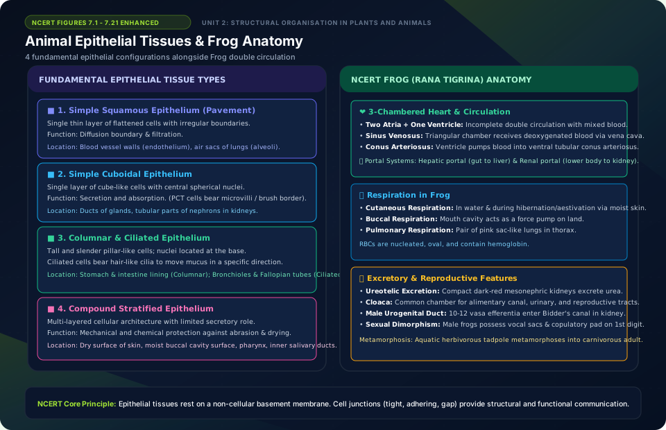
Animal Epithelial Tissues & Frog Anatomy
🚨 Examiner Traps & Common Misconceptions
✅ Reality: Tendons join skeletal Muscle to Bone (M-T-B). Ligaments join Bone to Bone (B-L-B).
✅ Reality: Cardiac muscle cells are short, branched, striated, and connected by intercalated discs with gap junctions that allow electrical signals to spread as a single synchronized syncytium.
💡 Memory Hack & Mnemonic
Connective Attachments: ‘Two Bones Linked by Ligament’ (Bone to Bone = Ligament); ‘Muscles Train Bones via Tendons’ (Muscle to Bone = Tendon).
⭐ 10/10 Active Recall Self-Check: Chapter 7
Test your mastery before closing this lecture note. Can you answer these without looking back?
- What are the primary diagnostic characteristics that define Structural Organisation in Animals in the NCERT syllabus?
- How do the physiological or anatomical structures covered in this chapter adapt the organism to its environment?
- What are the most common trick questions examiners create based on the exceptions in this chapter?
- Can you state the core mnemonic formula for remembering the sequential stages or components described above?
⭐ 10/10 Active Recall Self-Check: Chapter 7
Test your mastery before closing this lecture note. Can you answer these without looking back?
- What are the primary diagnostic characteristics that define Structural Organisation in Animals in the NCERT syllabus?
- How do the physiological or anatomical structures covered in this chapter adapt the organism to its environment?
- What are the most common trick questions examiners create based on the exceptions in this chapter?
- Can you state the core mnemonic formula for remembering the sequential stages or components described above?