
6.1 Meristematic Tissues: Apical, Intercalary & Lateral Meristems #
Plant anatomy explores the internal microscopic tissue organization of plant organs. A tissue is a group of cells having a common origin and performing a specific function. Plant tissues are broadly divided into two major groups: meristematic tissues (dividing cells) and permanent tissues (mature differentiated cells).
Growth in plants is largely restricted to specialized regions of active cell division called meristems. NCERT classifies meristems based on their position in the plant body:
- Apical Meristems: Located at the tips of roots and shoots, producing primary tissues that elongate the plant axis (Root Apical Meristem – RAM, and Shoot Apical Meristem – SAM). When shoot apical meristems produce leaves and stem elongation, cells ‘left behind’ form axillary buds in the axils of leaves capable of forming branches or flowers.
- Intercalary Meristems: Meristems occurring between mature tissues, notably at the base of internodes in grasses. They regenerate plant parts removed by grazing herbivores. Both apical and intercalary meristems are primary meristems because they appear early in plant life and contribute to the formation of the primary plant body.
- Lateral Meristems (Secondary Meristems): Cylindrical meristems that occur in the mature regions of roots and shoots of gymnosperms and dicots. They appear later than primary meristem and divide to produce secondary growth (increasing girth/thickness). Examples: Fascicular vascular cambium, Interfascicular cambium, and Cork cambium (phellogen).
6.2 Permanent Tissues: Simple (Parenchyma, Collenchyma, Sclerenchyma) & Complex (Xylem, Phloem) #
Cells produced by primary and secondary meristems lose the ability to divide and become specialized; these are permanent or mature tissues.
- Simple Permanent Tissues (Made of one cell type):
- Parenchyma: Living, isodiametric cells with thin cellulosic walls and small intercellular spaces. Functions: Photosynthesis (chlorenchyma), nutrient storage, and secretion.
- Collenchyma: Living cells with localized pectin, cellulose, and hemicellulose thickenings at the corners; intercellular spaces are absent. Occurs as a hypodermis below epidermis in dicot stems. Provides mechanical flexibility and tensile strength to growing young stems and petioles.
- Sclerenchyma: Dead cells with thick, highly lignified secondary walls and narrow lumens. Consists of elongated fibers (bast fibers) and round/oval sclereids (stone cells found in the pulp of guava, pear, sapota, and seed coats of legumes). Provides mechanical support.
- Complex Permanent Tissues (Made of multiple cell types working together):
- Xylem (Water & Mineral Conduction): Composed of four elements:
- Tracheids: Elongated, dead, tube-like lignified cells with tapering ends and bordered pits.
- Vessels: Long cylindrical tubes made of vessel members with perforated end walls. Presence of vessels is a diagnostic hallmark of angiosperms (gymnosperms lack vessels).
- Xylem Fibers: Dead, highly thickened fibers providing rigid support.
- Xylem Parenchyma: The only living element in xylem; stores starch and fats and conducts water radially (ray parenchyma).
- Phloem (Food Translocation): Composed of four elements:
- Sieve Tube Elements: Long, tube-like living cells arranged longitudinally with perforated end walls called sieve plates. Mature sieve tube elements possess peripheral cytoplasm and a large vacuole but lack a nucleus! Their functions are controlled by the nucleus of adjacent companion cells.
- Companion Cells: Specialized parenchymatous cells connected to sieve tubes by pit fields. Maintain pressure gradient in sieve tubes.
- Phloem Parenchyma: Living cells storing resins, latex, and mucilage (absent in most monocots).
- Phloem Fibers (Bast Fibers): Sclerenchymatous dead fibers providing mechanical support (e.g., jute, flax, and hemp fibers used commercially).
- Xylem (Water & Mineral Conduction): Composed of four elements:
6.3 Tissue Systems: Epidermal, Ground (Cortex, Pericycle, Pith) & Vascular System #
Based on their location and functional specialization, all plant tissues in a vegetative organ are organized into three distinct tissue systems:
- Epidermal Tissue System: Outermost protective skin of the plant body. Consists of epidermal cells, cuticular wax coating, stomata, and appendages. Stomata regulate transpiration and gas exchange; each stoma consists of two bean-shaped guard cells (dumbbell-shaped in grasses) containing chloroplasts, surrounding a stomatal aperture. Epidermal hairs on stems are multicellular and branched/unbranched, called trichomes (prevent water loss and secrete substances), whereas root hairs are unicellular elongations.
- Ground (Fundamental) Tissue System: Forms the main bulk of the plant body between epidermis and vascular bundles. Consists of parenchyma, collenchyma, and sclerenchyma. In roots and stems, it differentiates into hypodermis, general cortex, endodermis, pericycle, medullary rays, and pith. In leaves, the ground tissue consists of thin-walled, chloroplast-rich photosynthetic cells called mesophyll.
- Vascular Tissue System: Consists of complex conducting tissues—xylem and phloem arranged in vascular bundles:
- Radial Vascular Bundles: Xylem and phloem arranged alternately along different radii. Characteristic of all roots (dicot and monocot).
- Conjoint Vascular Bundles: Xylem and phloem jointly situated on the same radius. Characteristic of stems and leaves.
- Conjoint Open: Cambium is present between xylem and phloem, capable of secondary growth (characteristic of dicot stems).
- Conjoint Closed: Cambium is completely absent; no secondary growth occurs (characteristic of monocot stems).
6.4 Anatomy of Dicotyledonous vs Monocotyledonous Root #
Examining transverse sections (T.S.) of dicotyledonous (e.g., Sunflower) and monocotyledonous (e.g., Maize) roots reveals critical internal anatomical distinctions:
| Anatomical Zone | Dicotyledonous Root (Sunflower) | Monocotyledonous Root (Maize) |
|---|---|---|
| Epiblema (Piliferous Layer) | Uniseriate epidermis bearing unicellular root hairs; cuticle absent. | Uniseriate epidermis with unicellular root hairs. |
| Endodermis | Single layer of barrel-shaped cells with suberin-impregnated waterproof bands called Casparian strips on radial and tangential walls. | Casparian strips present; passage cells opposite protoxylem vessels. |
| Pericycle | Thick-walled parenchymatous cells giving rise to lateral roots and part of the vascular cambium. | Gives rise only to lateral roots (no secondary growth). |
| Vascular Bundles | Radial; Diarch to Hexarch (2 to 6 xylem and phloem patches); Xylem is exarch (protoxylem toward periphery, metaxylem toward center). | Radial; Polyarch (more than 6 xylem patches, often 8 to 20); Xylem is exarch. |
| Pith | Small or completely inconspicuous/absent. | Large, well-developed and parenchymatous. |
| Secondary Growth | Occurs actively via vascular cambium and cork cambium. | Completely absent. |
6.5 Anatomy of Dicotyledonous vs Monocotyledonous Stem #
In stems, the orientation of primary xylem is endarch (protoxylem lies toward the center/pith, while metaxylem lies toward the periphery). A comparison between dicot and monocot stem anatomy highlights fundamental structural differences:
- Dicotyledonous Stem (e.g., Sunflower):
- Epidermis: Covered by a protective cuticular layer with trichomes and stomata.
- Cortex: Clearly stratified into three zones: outer collenchymatous hypodermis (mechanical support), middle parenchymatous general cortex, and innermost endodermis (rich in starch grains, termed the starch sheath).
- Pericycle: Present on the inner side of endodermis above vascular bundles as semi-lunar patches of sclerenchyma.
- Vascular Bundles: Arranged in a neat broken ring (hallmark of dicot stem); each bundle is conjoint, collateral, open (with cambium between xylem and phloem), with endarch xylem.
- Pith: Large, central parenchymatous pith with prominent medullary rays running radially.
- Monocotyledonous Stem (e.g., Maize):
- Hypodermis: Composed of sclerenchymatous cells (providing rigid mechanical strength).
- Ground Tissue: Huge, continuous mass of undifferentiated parenchyma; there is no demarcation into cortex, endodermis, pericycle, or pith!
- Vascular Bundles: Scattered throughout ground tissue; smaller bundles are concentrated toward periphery, while larger bundles lie toward center. Each bundle is surrounded by a sclerenchymatous bundle sheath. Bundles are conjoint, collateral, and closed (lacking cambium). Phloem parenchyma is absent, and water-containing cavities (lysigenous cavities) are present within the vascular bundles.
6.6 Anatomy of Dorsiventral (Dicot) vs Isobilateral (Monocot) Leaf #
Leaves exhibit remarkable internal histology adapted for light harvesting, carbon dioxide uptake, and minimal transpirational water loss:
| Anatomical Zone | Dorsiventral Leaf (Dicot Leaf) | Isobilateral Leaf (Monocot Leaf) |
|---|---|---|
| Stomatal Distribution | Hypostomatic: Stomata are significantly more numerous on the lower (abaxial) epidermis than on upper (adaxial) epidermis. | Amphistomatic: Stomata are distributed equally on both adaxial and abaxial epidermises. |
| Mesophyll Differentiation | Differentiated into two distinct layers:
| Undifferentiated mesophyll: Composed of uniform isodiametric cells with small intercellular spaces; no distinction into palisade and spongy parenchyma. |
| Bulliform Cells | Absent. | Present! Large, empty, colorless specialized adaxial epidermal cells in grasses. When turgid, leaf blade unfolds; when flaccid under water stress, leaves curl inward to minimize water loss. |
| Venation & Vascular Size | Vascular bundles vary in size depending on reticulate venation (largest in midrib). | Vascular bundles are nearly uniform in size along parallel veins. |
6.7 Secondary Growth: Vascular Cambium, Cork Cambium (Phellogen), Heartwood vs Sapwood, Annual Rings #
Secondary Growth: Vascular Cambium, Cork Cambium (Phellogen), Heartwood vs Sapwood, Annual Rings is a core concept in this chapter. Understanding this requires analyzing its structural organization, biological mechanisms, and regulatory feedback pathways.
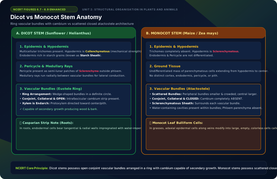
Dicot vs Monocot Stem Anatomy
🚨 Examiner Traps & Common Misconceptions
✅ Reality: Monocot stems have scattered closed vascular bundles with no cambium between xylem and phloem, so they do not exhibit normal secondary growth!
✅ Reality: Mature xylem vessel elements and tracheids are dead, hollow tubes with lignified walls designed for maximum fluid transmission without cytoplasmic resistance.
💡 Memory Hack & Mnemonic
Periderm Layers (Outside to Inside): ‘Phellem, Phellogen, Phelloderm’ = ‘Cork, Cork Cambium, Secondary Cortex’.
⭐ 10/10 Active Recall Self-Check: Chapter 6
Test your mastery before closing this lecture note. Can you answer these without looking back?
- What are the primary diagnostic characteristics that define Anatomy of Flowering Plants in the NCERT syllabus?
- How do the physiological or anatomical structures covered in this chapter adapt the organism to its environment?
- What are the most common trick questions examiners create based on the exceptions in this chapter?
- Can you state the core mnemonic formula for remembering the sequential stages or components described above?
⭐ 10/10 Active Recall Self-Check: Chapter 6
Test your mastery before closing this lecture note. Can you answer these without looking back?
- What are the primary diagnostic characteristics that define Anatomy of Flowering Plants in the NCERT syllabus?
- How do the physiological or anatomical structures covered in this chapter adapt the organism to its environment?
- What are the most common trick questions examiners create based on the exceptions in this chapter?
- Can you state the core mnemonic formula for remembering the sequential stages or components described above?