
2.1 Historical Systems: Two Kingdom to Five Kingdom Classification (R.H. Whittaker 1969) #
Since the dawn of civilization, humans have attempted to classify living organisms based on practical utility—food, shelter, and clothing. Aristotle was the earliest scientist to establish a scientific basis for classification, categorizing plants into simple morphological forms (trees, shrubs, and herbs) and animals into two groups: those with red blood (Enaima) and those without red blood (Anaima).
Later, Carolus Linnaeus developed the Two Kingdom System, dividing all life into Kingdom Plantae and Kingdom Animalia. However, this system failed to distinguish between eukaryotes and prokaryotes, unicellular and multicellular organisms, or photosynthetic green algae and non-photosynthetic fungi.
To overcome these major shortcomings, R.H. Whittaker (1969) proposed the groundbreaking Five Kingdom Classification. The primary criteria used by Whittaker to construct the five kingdoms include:
- Cell Structure: Prokaryotic (lacking membrane-bound nucleus) vs Eukaryotic.
- Body Organization: Unicellular vs Multicellular tissue/organ level.
- Mode of Nutrition: Autotrophic (photosynthetic/chemosynthetic) vs Heterotrophic (absorptive in Fungi, ingestive/holozoic in Animals).
- Reproduction: Asexual vs sexual reproductive strategies.
- Phylogenetic Relationships: Evolutionary lineages and ancestral connections.
Whittaker’s five kingdoms are: Monera, Protista, Fungi, Plantae, and Animalia.
2.2 Kingdom Monera: Archaebacteria, Eubacteria, Cyanobacteria & Mycoplasma #
Bacteria are the sole members of Kingdom Monera. They are the most abundant microorganisms on Earth, thriving in extreme habitats like deep ocean hydrothermal vents, hot sulfur springs, snow, and deserts where other life forms cannot survive.
Kingdom Monera is divided into two major evolutionary groups:
- Archaebacteria (Primitive Bacteria): These ancient organisms survive the most hostile conditions on Earth due to their unique cell wall structure (lacking peptidoglycan) and branched ether-linked membrane lipids.
- Halophiles: Live in extremely high saline environments (e.g., salt lakes).
- Thermoacidophiles: Thrive in boiling, highly acidic sulfur hot springs (temperature ~80°C, pH ~2).
- Methanogens: Strict anaerobes living in marshy areas and in the gut of ruminant animals (cows and buffaloes), responsible for producing biogas (methane) from dung.
- Eubacteria (‘True Bacteria’): Characterized by a rigid peptidoglycan cell wall and, if motile, a flagellum composed of flagellin protein.
- Cyanobacteria (Blue-Green Algae): Unicellular, colonial, or filamentous photosynthetic autotrophs containing Chlorophyll a (identical to green plants). Many fix atmospheric nitrogen inside specialized thick-walled, non-photosynthetic anaerobic cells called heterocysts (e.g., Nostoc and Anabaena).
- Chemosynthetic Autotrophs: Oxidize inorganic substances like nitrates, nitrites, and ammonia to release ATP, recycling nitrogen, phosphorus, iron, and sulfur.
- Heterotrophic Bacteria: The most abundant in nature, acting as vital decomposers. They produce curd from milk (Lactobacillus), yield antibiotics, fix nitrogen in legume roots (Rhizobium), or cause diseases such as cholera (Vibrio cholerae), typhoid (Salmonella typhi), and tetanus (Clostridium tetani).
- Mycoplasma: Organisms that completely lack a cell wall. They are the smallest known living cells (0.3 µm) and can survive without oxygen. Many are pathogenic in animals and plants.
2.3 Kingdom Protista: Chrysophytes, Dinoflagellates, Euglenoids, Slime Moulds & Protozoans #
Kingdom Protista brings together all single-celled eukaryotes. The boundaries of this kingdom are fluid because what is a photosynthetic plant-like protist to one biologist may be an algal cell to another. Protistan cells possess a well-defined nucleus and membrane-bound organelles. They are primarily aquatic and reproduce asexually and sexually via zygote formation.
NCERT classifies Kingdom Protista into five major groups:
| Protist Group | Diagnostic Features & Cell Coverings | Key Examples & Ecological Roles |
|---|---|---|
| Chrysophytes | Diatoms and Golden algae (Desmids). Cell walls form two thin overlapping shells fitting like a soapbox, embedded with indestructible silica. | Diatomaceous earth used in polishing and filtration. Diatoms are the chief ‘producers’ in the oceans. |
| Dinoflagellates | Mostly marine, photosynthetic with stiff cellulose plates on cell walls. Possess two flagella: one longitudinal and one transverse in a furrow. | Gonyaulax undergoes rapid multiplication causing ‘red tides’ and releases toxins (saxitoxin) killing fish. |
| Euglenoids | Freshwater organisms lacking cell walls; wrapped in a protein-rich flexible layer called pellicle. Possess one short and one long flagellum. | Euglena: Mixotrophic nutrition (photosynthetic in sunlight, predatory heterotroph in darkness). |
| Slime Moulds | Saprophytic protists moving along decaying twigs engulfing organic matter. Under favorable conditions, they form an aggregate called plasmodium. | In unfavorable conditions, plasmodium differentiates to form fruiting bodies bearing spores with true resistant walls dispersed by air currents. |
| Protozoans | Heterotrophic unicellular predators or parasites: Amoeboid (pseudopodia), Flagellated, Ciliated (gullet and rows of cilia), and Sporozoans (infectious spore-like stage). | Amoeba, Trypanosoma (sleeping sickness), Paramoecium (ciliated), Plasmodium (malarial parasite). |
2.4 Kingdom Fungi: Phycomycetes, Ascomycetes, Basidiomycetes & Deuteromycetes #
Kingdom Fungi constitutes a unique group of heterotrophic, achlorophyllous, eukaryotic organisms. Their bodies consist of long, slender, thread-like structures called hyphae. The network of hyphae is known as mycelium. Hyphae may be continuous tubes filled with multinucleated cytoplasm (coenocytic) or divided by cross-walls called septa.
The fungal cell wall is composed of chitin and polysaccharides. Fungi obtain nutrients by absorbing soluble organic matter from dead substrates (saprophytes), living plants and animals (parasites), or as symbionts (lichens with algae, and mycorrhizae with roots of higher plants).
NCERT classifies Kingdom Fungi into four major classes based on the morphology of the mycelium, mode of spore formation, and fruiting bodies:
- Phycomycetes (Algal Fungi): Found in aquatic habitats and on decaying wood in moist places. Mycelium is aseptate and coenocytic. Asexual reproduction occurs by motile zoospores or non-motile aplanospores formed endogenously inside a sporangium. Sexual spores are zygospores formed by fusion of gametes (isogamous or oogamous). Examples: Mucor, Rhizopus (bread mould), and Albugo (parasitic white rust on mustard).
- Ascomycetes (Sac Fungi): Mostly multicellular (e.g., Penicillium), rarely unicellular (e.g., Yeast, Saccharomyces). Mycelium is branched and septate. Asexual spores are conidia produced exogenously on special hyphae called conidiophores. Sexual spores are ascospores produced endogenously in sac-like structures called asci (singular: ascus) packed in fruiting bodies called ascocarps. Examples: Aspergillus, Claviceps, Neurospora (used extensively in genetics), and edible morels and truffles.
- Basidiomycetes (Club Fungi): Mushrooms, bracket fungi, and puffballs. Mycelium is branched and septate. Asexual spores are generally not found, but vegetative reproduction by fragmentation is common. Sex organs are absent, but plasmogamy occurs by fusion of two somatic cells to form a dikaryotic mycelium, which produces basidia bearing 4 basidiospores exogenously on fruiting bodies called basidiocarps. Examples: Agaricus (common edible mushroom), Ustilago (smut), and Puccinia (wheat rust fungus).
- Deuteromycetes (Fungi Imperfecti): Commonly called imperfect fungi because only their asexual or vegetative phases are known. When sexual stages of these fungi are discovered, they are transferred to Ascomycetes or Basidiomycetes. Mycelium is septate and branched. They reproduce asexually only by conidia. Many are decomposers of litter aiding mineral cycling. Examples: Alternaria, Colletotrichum, and Trichoderma.
2.5 Acellular Entities: Viruses, Viroids, Prions & Symbiotic Lichens #
Whittaker’s Five Kingdom Classification did not include acellular entities like viruses, viroids, and prions, nor did it include composite symbiotic organisms like lichens.
- Viruses: Non-cellular entities characterized by an inert crystalline protein structure outside the living host cell. Once they infect a host cell, they take over the cell’s metabolic machinery to replicate themselves, killing the host. A virus consists of genetic material enclosed in a protective protein coat called a capsid (composed of subunit capsomeres). A virus contains either RNA or DNA, never both. Plant viruses usually have single-stranded RNA (ssRNA); animal viruses have ssRNA, dsRNA, or dsDNA; and bacterial viruses (bacteriophages) usually possess double-stranded DNA (dsDNA). Key scientists: D.J. Ivanowsky (1892) discovered tobacco mosaic virus; M.W. Beijerinek (1898) coined the term Contagium vivum fluidum (infectious living fluid); and W.M. Stanley (1935) crystallized viruses showing capsids are mostly proteins.
- Viroids: Discovered by T.O. Diener in 1971 as infectious agents smaller than viruses. Viroids consist of free, single-stranded RNA of low molecular weight, completely lacking a protein capsid. They cause Potato spindle tuber disease.
- Prions: Abnormally folded proteins capable of transmitting infectious neurological diseases without any genetic nucleic acid. They cause Bovine Spongiform Encephalopathy (BSE, or mad cow disease) in cattle and its human variant, Cr-Jacob disease (CJD).
- Lichens: Symbiotic associations between an alga (phycobiont, which is autotrophic and prepares food) and a fungus (mycobiont, which provides shelter, absorbs minerals, and anchors moisture). Lichens are pioneer colonizers on bare rock and act as sensitive pollution indicators because they never grow in sulfur dioxide-polluted environments.
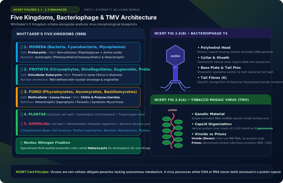
Five Kingdoms, Bacteriophage & TMV Architecture
🚨 Examiner Traps & Common Misconceptions
✅ Reality: Viruses are non-cellular inert crystalline structures outside their living host. They contain only a protein capsid and genetic material (RNA or DNA, never both!).
✅ Reality: T.O. Diener discovered viroids in 1971: they are free infectious RNA molecules of low molecular weight, lacking any protein coat completely (causing Potato Spindle Tuber disease).
💡 Memory Hack & Mnemonic
Four Fungal Classes: ‘Please Ask Bob Down’ (Phycomycetes, Ascomycetes, Basidiomycetes, Deuteromycetes – Fungi Imperfecti).
⭐ 10/10 Active Recall Self-Check: Chapter 2
Test your mastery before closing this lecture note. Can you answer these without looking back?
- What are the primary diagnostic characteristics that define Biological Classification in the NCERT syllabus?
- How do the physiological or anatomical structures covered in this chapter adapt the organism to its environment?
- What are the most common trick questions examiners create based on the exceptions in this chapter?
- Can you state the core mnemonic formula for remembering the sequential stages or components described above?
⭐ 10/10 Active Recall Self-Check: Chapter 2
Test your mastery before closing this lecture note. Can you answer these without looking back?
- What are the primary diagnostic characteristics that define Biological Classification in the NCERT syllabus?
- How do the physiological or anatomical structures covered in this chapter adapt the organism to its environment?
- What are the most common trick questions examiners create based on the exceptions in this chapter?
- Can you state the core mnemonic formula for remembering the sequential stages or components described above?