
19.1 Endocrine Glands vs Exocrine Glands; Definition of Hormones (Non-nutrient chemical messengers in trace amounts) #
While the neural system provides rapid point-to-point bioelectrical coordination, neural fibers do not innervate all cells of the body, and cellular functions require continuous, long-term chemical regulation. This coordination is provided by the Endocrine System via chemical messengers called hormones.
Endocrine Glands: Ductless glands that secrete their products directly into the circulating blood. NCERT defines hormones as: “Non-nutrient chemicals which act as intercellular messengers and are produced in trace amounts.”
The classical endocrine glands in humans include the Hypothalamus, Pituitary, Pineal, Thyroid, Parathyroid, Thymus, Adrenal, Pancreas, and Gonads (Testes and Ovaries). In addition, organized endocrine tissues are found in the gastrointestinal tract, liver, kidney, and heart.
19.2 Hypothalamus: Releasing Hormones (GnRH) & Inhibiting Hormones (Somatostatin); Posterior Pituitary Connections #
The Hypothalamus forms the basal part of the diencephalon (forebrain) and acts as the supreme master coordinating bridge between the nervous system and the endocrine system. It contains clusters of neurosecretory cells called nuclei that synthesize and secrete two classes of regulatory neurohormones:
- Releasing Hormones: Stimulate the secretion of anterior pituitary hormones (e.g., Gonadotropin-Releasing Hormone [GnRH] stimulates the pituitary to release LH and FSH).
- Inhibiting Hormones: Inhibit the secretion of pituitary hormones (e.g., Somatostatin inhibits the secretion of Growth Hormone from the pituitary).
These hypothalamic hormones reach the anterior pituitary through a specialized hypophyseal portal circulatory system. In contrast, the posterior pituitary is under direct axonal neural regulation from the hypothalamus.
19.3 Pituitary Gland (Hypophysis): Adenohypophysis (GH, PRL, TSH, ACTH, LH, FSH) & Neurohypophysis (Oxytocin, Vasopressin/ADH) #
The Pituitary Gland (Hypophysis) is located in a bony cavity of the sphenoid bone called the sella turcica, attached to the hypothalamus by a vascular stalk (infundibulum). It is divided anatomically into two distinct lobes:
- Adenohypophysis (Anterior Pituitary): Consists of two portions: Pars distalis and Pars intermedia.
- Growth Hormone (GH): Stimulates body growth and protein synthesis. Hypersecretion in childhood causes Gigantism; hypersecretion in adults causes disfiguring bone enlargement called Acromegaly; hyposecretion in childhood results in Pituitary Dwarfism.
- Prolactin (PRL): Regulates mammary gland growth and lactation (milk production).
- Thyroid-Stimulating Hormone (TSH): Stimulates thyroid gland to synthesize and release thyroid hormones.
- Adrenocorticotropic Hormone (ACTH): Stimulates adrenal cortex to secrete glucocorticoids.
- Luteinizing Hormone (LH) & Follicle-Stimulating Hormone (FSH): Known collectively as Gonadotropins. In males, LH stimulates Leydig cells to secrete androgens (testosterone), and FSH stimulates spermatogenesis. In females, LH induces ovulation of mature Graafian follicles and maintains the corpus luteum; FSH stimulates ovarian follicular growth.
- Melanocyte-Stimulating Hormone (MSH): Secreted by Pars intermedia; acts on melanocytes to regulate skin pigmentation.
- Neurohypophysis (Pars Nervosa / Posterior Pituitary): Does not synthesize hormones itself! It stores and releases two hormones synthesized in hypothalamic neurons:
- Oxytocin: Stimulates vigorous smooth muscle contractions of the uterus during childbirth (labor hormone) and milk ejection from mammary glands (‘milk let-down hormone’).
- Vasopressin / Antidiuretic Hormone (ADH): Stimulates water and electrolyte reabsorption from the DCT and collecting ducts of kidneys, preventing water loss (diuresis). Hyposecretion of ADH results in Diabetes Insipidus (excretion of huge volumes of dilute urine and severe thirst, without glucose in urine).
19.4 Pineal Gland: Melatonin & Regulation of 24-Hour Circadian Diurnal Rhythm #
The Pineal Gland is a tiny, pinecone-shaped endocrine gland located on the dorsal side of the forebrain. It secretes the indoleamine hormone Melatonin.
Melatonin plays a critical regulatory role in maintaining the body’s 24-hour diurnal (circadian) rhythm:
- Regulates the normal sleep-wake cycle (melatonin levels surge in darkness inducing sleep, and drop with morning light).
- Regulates core body temperature rhythm.
- Influences metabolism, skin pigmentation, menstrual cycle, and our immune defense capabilities.
19.5 Thyroid Gland: T3, T4 (Thyroxine) & Thyrocalcitonin (TCT); Hypothyroidism (Goitre, Cretinism) vs Hyperthyroidism (Graves’ Disease) #
The Thyroid Gland consists of two vascular lobes situated on either side of the trachea in the neck, connected by a thin median flap of connective tissue called the isthmus. It is composed of microscopic spherical thyroid follicles surrounded by stromal tissues. Follicular cells require dietary iodine to synthesize two iodinated hormones: Triiodothyronine (T3) and Tetraiodothyronine / Thyroxine (T4):
- Physiological Functions of Thyroid Hormones: Regulate the Basal Metabolic Rate (BMR), stimulate red blood cell formation (erythropoiesis), control metabolism of carbohydrates, proteins, and fats, and maintain water and electrolyte balance.
- Thyrocalcitonin (TCT): A peptide hormone secreted by parafollicular C-cells of the thyroid. TCT is a hypocalcemic hormone that lowers blood calcium levels by promoting calcium deposition in bones.
- Thyroid Disorders:
- Hypothyroidism: Deficiency of dietary iodine causes enlargement of the thyroid gland, commonly called Simple Goitre. Hypothyroidism during pregnancy results in defective fetal development leading to stunted growth, mental retardation, low IQ, abnormal skin, and deaf-mutism in children (Cretinism). In adult women, it causes irregular menstrual cycles.
- Hyperthyroidism: Over-secretion of thyroid hormones due to thyroid nodules or cancer. A common form is Exophthalmic Goitre (Graves’ Disease), an autoimmune disorder characterized by thyroid enlargement, protruding eyeballs (exophthalmos), increased BMR, and severe weight loss.
19.6 Parathyroid Glands: Parathyroid Hormone (PTH / Hypercalcemic Hormone) Antagonizing TCT #
In humans, four tiny Parathyroid Glands are present on the back side of the thyroid gland (one pair on each thyroid lobe). They secrete a peptide hormone called Parathyroid Hormone (PTH), also known as Collip’s hormone.
PTH is an essential hypercalcemic hormone that increases blood Ca2+ levels:
- Stimulates bone resorption (demineralization) by activating osteoclasts to dissolve bone matrix and release Ca2+ into blood.
- Stimulates reabsorption of Ca2+ by the renal tubules in kidneys.
- Increases Ca2+ absorption from digested food in the intestine.
Calcium Homeostasis: PTH and TCT act as direct antagonists to precisely regulate and maintain blood calcium levels (~9–11 mg/dL) vital for muscle contraction, nerve transmission, and blood clotting.
19.7 Thymus Gland: Thymosins & Differentiation of T-Lymphocytes (Cell-Mediated Immunity) #
The Thymus Gland is a lobular primary lymphoid organ situated between the lungs behind the sternum on the ventral side of the aorta. It secretes peptide hormones called Thymosins.
- Thymosins play a major role in the differentiation and maturation of T-lymphocytes, which provide Cell-Mediated Immunity (CMI).
- Thymosins also promote antibody production, providing Humoral Immunity.
- Age-related Atrophy: The thymus is prominent at birth but progressively degenerates and shrinks with aging. Consequently, production of thymosins drops severely, leading to the characteristic weakening of the immune system in elderly individuals.
19.8 Adrenal Gland: Adrenal Cortex (Aldosterone, Cortisol, Androgens) & Adrenal Medulla (Adrenaline / Epinephrine 3F Emergency Hormone) #
The human body possesses a pair of Adrenal (Suprarenal) Glands, one located atop each kidney. An adrenal gland is composed of two distinct structural and functional endocrine tissues:
- Adrenal Medulla (Inner Core): Derived from embryonic neural crest cells; secretes two catecholamine hormones: Adrenaline (Epinephrine) and Noradrenaline (Norepinephrine).
Commonly called the Emergency Hormones or Hormones of Fight or Flight (3F Hormones). Released rapidly in response to stress, fear, or emergency conditions. They cause pupillary dilation, piloerection (raising of body hairs), sweating, increase heart rate, cardiac contractility, respiratory rate, and stimulate glycogenolysis and lipolysis to raise blood glucose concentrations.
- Adrenal Cortex (Outer Shell): Divided into three distinct cellular zones (from outside in):
- Zona glomerulosa (Outer): Secretes Mineralocorticoids, primarily Aldosterone. Aldosterone acts on renal DCT and collecting ducts to stimulate active reabsorption of Na+ and water, and excretion of K+ and phosphate, maintaining blood volume and blood pressure.
- Zona fasciculata (Middle): Secretes Glucocorticoids, primarily Cortisol. Cortisol stimulates gluconeogenesis, lipolysis, and proteolysis; produces anti-inflammatory reactions and suppresses the immune response; and stimulates RBC production.
- Zona reticularis (Inner): Secretes small amounts of Androgenic steroids that stimulate the development of axial, pubic, and facial hair during puberty.
19.9 Pancreas (Islets of Langerhans): Glucagon (Alpha cells / Hyperglycemic) & Insulin (Beta cells / Hypoglycemic; Diabetes Mellitus) #
The Pancreas is a composite heterocrine (dual) gland acting as both an exocrine gland (secreting pancreatic digestive enzymes) and an endocrine gland. The endocrine pancreas consists of approximately 1 to 2 million microscopic cell clusters called the Islets of Langerhans, representing only 1–2% of pancreatic tissue:
- Alpha (α) Cells (~25%): Secrete the peptide hormone Glucagon.
- Glucagon is a hyperglycemic hormone that raises blood glucose levels.
- Acts primarily on liver hepatocytes, stimulating glycogenolysis (breakdown of glycogen into glucose) and gluconeogenesis.
- Reduces cellular glucose uptake and utilization.
- Beta (β) Cells (~70%): Secrete the peptide hormone Insulin.
- Insulin is a hypoglycemic hormone that lowers blood glucose levels.
- Acts on hepatocytes and adipocytes, rapidly stimulating cellular glucose uptake and utilization.
- Stimulates glycogenesis (conversion of glucose into glycogen).
Prolonged hyposecretion or cellular resistance to insulin results in a complex metabolic disorder called Diabetes Mellitus. It is characterized by chronic hyperglycemia (high blood sugar), glycosuria (loss of glucose in urine), and the formation of toxic breakdown products called ketone bodies (diabetic ketoacidosis). Diabetic patients are successfully managed with insulin therapy.
19.10 Gonadal Hormones: Testis (Testosterone / Androgens) & Ovary (Estrogen & Progesterone) #
The male and female reproductive gonads act as vital endocrine glands producing steroid sex hormones:
- Testes (Male Gonads): Located in the extra-abdominal scrotum. Interstitial spaces between seminiferous tubules contain endocrine Leydig Cells (Interstitial Cells) that synthesize and secrete steroid hormones called Androgens (principally Testosterone):
- Regulates development, maturation, and functions of male accessory sex organs (epididymis, vas deferens, seminal vesicles, prostate).
- Stimulates male secondary sexual characteristics (deep voice, beard, mustache, aggressive masculine muscular build).
- Stimulates spermatogenesis and exerts an anabolic effect on protein and carbohydrate metabolism.
- Ovaries (Female Gonads): Located in the lower abdominal pelvic cavity. The ovary produces two groups of steroid hormones: Estrogen and Progesterone:
- Estrogen: Synthesized and secreted by growing ovarian follicles. Stimulates growth of female secondary sex organs, development of ovarian follicles, feminine secondary sexual characters (high pitch voice, mammary gland development), and feminine sexual behavior.
- Progesterone: Secreted by the corpus luteum (the ruptured follicle post-ovulation). Known as the pregnancy hormone: supports pregnancy, stimulates thickening of the uterine endometrium, and acts on mammary glands to stimulate formation of alveoli (sacs storing milk) and milk secretion.
19.11 Hormones of Non-Endocrine Tissues: Heart (ANF), Kidney (Erythropoietin) & Gastrointestinal Tract (Gastrin, Secretin, CCK, GIP) #
In addition to organized endocrine glands, endocrine cells are present in other non-endocrine vital organs that secrete critical peptide hormones:
- Heart: The muscular atrial wall of the heart secretes a peptide hormone called Atrial Natriuretic Factor (ANF) when blood pressure increases. ANF causes vasodilation, lowering blood pressure.
- Kidney: The juxtaglomerular cells of the kidney secrete a peptide hormone called Erythropoietin (EPO), which stimulates the bone marrow to produce red blood cells (erythropoiesis) in response to hypoxia.
- Gastrointestinal Tract (GIT): Endocrine cells scattered along the mucosa of the stomach and intestine secrete four major peptide hormones:
- Gastrin: Acts on gastric glands to stimulate secretion of hydrochloric acid (HCl) and pepsinogen.
- Secretin: Acts on the exocrine pancreas to stimulate secretion of water and bicarbonate ions.
- Cholecystokinin (CCK): Acts on both gallbladder (stimulating contraction to release bile salts) and pancreas (stimulating secretion of pancreatic enzymes).
- Gastric Inhibitory Peptide (GIP): Inhibits gastric motility and gastric secretions.
19.12 Mechanisms of Hormone Action: Peptide Hormones via Membrane Receptors & Second Messengers (cAMP, IP3, Ca2+) vs Lipid-Soluble Steroid Hormones via Intracellular Nuclear Receptors #
Hormones exert their biological effects on target tissues by binding specifically to high-affinity protein molecules called Hormone Receptors located only in target tissues. Based on their chemical nature and mechanism of action, hormones are divided into two fundamental classes:
- Water-Soluble Peptide / Protein / Amine Hormones: (e.g., Insulin, Glucagon, Pituitary hormones, Epinephrine).
These polar hormones cannot diffuse across the lipid bilayer of the target cell membrane. Instead, they bind to Membrane-Bound Receptors on the cell surface:
- Hormone binds to surface receptor, forming a Hormone-Receptor Complex.
- This activates membrane G-proteins, stimulating an amplifier enzyme (such as adenylate cyclase).
- Generates Second Messengers inside the cell (e.g., cyclic AMP [cAMP], Inositol Triphosphate [IP3], or Calcium ions [Ca2+]).
- Second messengers trigger an enzymatic phosphorylation cascade, producing rapid cellular biochemical and physiological responses. The hormone itself never enters the cell!
- Lipid-Soluble Steroid & Thyroid Hormones: (e.g., Estrogen, Progesterone, Testosterone, Cortisol, Aldosterone, and Thyroxine).
Because they are lipid-soluble, these hormones diffuse freely across the target cell membrane and bind to Intracellular Receptors (mostly nuclear receptors):
- Hormone enters the nucleus and binds to its specific intracellular receptor.
- The Hormone-Receptor complex binds directly to specific regulatory DNA sequences on the chromosome.
- This directly alters gene expression and transcription of specific mRNAs, directing protein synthesis and producing long-term physiological and developmental changes.
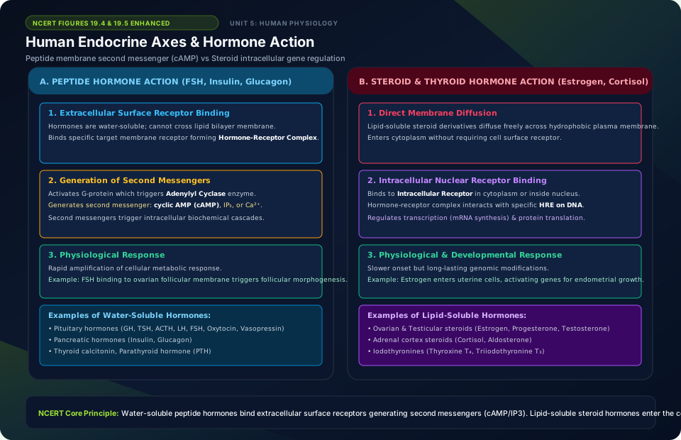
Human Endocrine Axes & Hormone Action
🚨 Examiner Traps & Common Misconceptions
✅ Reality: They are synthesized by hypothalamic neurosecretory neurons and only STORED and released by the neurohypophysis!
✅ Reality: Insulin is strictly HYPOGLYCEMIC: it lowers blood glucose by accelerating cellular glucose uptake and glycogenesis in liver and muscles.
💡 Memory Hack & Mnemonic
Calcium Regulation Hormones: ‘CalciTONin TONES down calcium (lowers blood Ca2+); PTH PUMPS UP calcium (raises blood Ca2+)’.
⭐ 10/10 Active Recall Self-Check: Chapter 19
Test your mastery before closing this lecture note. Can you answer these without looking back?
- What are the primary diagnostic characteristics that define Chemical Coordination and Integration in the NCERT syllabus?
- How do the physiological or anatomical structures covered in this chapter adapt the organism to its environment?
- What are the most common trick questions examiners create based on the exceptions in this chapter?
- Can you state the core mnemonic formula for remembering the sequential stages or components described above?
⭐ 10/10 Active Recall Self-Check: Chapter 19
Test your mastery before closing this lecture note. Can you answer these without looking back?
- What are the primary diagnostic characteristics that define Chemical Coordination and Integration in the NCERT syllabus?
- How do the physiological or anatomical structures covered in this chapter adapt the organism to its environment?
- What are the most common trick questions examiners create based on the exceptions in this chapter?
- Can you state the core mnemonic formula for remembering the sequential stages or components described above?