
18.1 Neural System Organization: Central Nervous System (CNS) & Peripheral Nervous System (PNS: Somatic & Autonomic) #
As organisms evolved complex multicellular organ systems, maintaining homeostasis required rapid coordination. Coordination is the process through which two or more organs interact and complement the functions of one another. In animals, the neural system provides an organized network of point-to-point connections for rapid coordination via bioelectrical impulses.
The human neural system is divided into two primary structural divisions:
- Central Nervous System (CNS): Comprises the Brain and the Spinal Cord; the master control and processing site for information.
- Peripheral Nervous System (PNS): Comprises all the nerves of the body associated with the CNS (12 pairs of cranial nerves and 31 pairs of spinal nerves). Nerve fibers are of two types:
- Afferent (Sensory) Fibers: Transmit sensory impulses from tissues/organs to the CNS.
- Efferent (Motor) Fibers: Transmit regulatory motor impulses from the CNS to peripheral target effector organs.
- Somatic Neural System (SNS): Relays impulses from CNS to voluntary skeletal muscles.
- Autonomic Neural System (ANS): Transmits impulses from CNS to involuntary visceral organs and smooth muscles. Consists of the Sympathetic division (prepares body for fight-or-flight emergencies) and Parasympathetic division (promotes rest-and-digest recovery).
18.2 Neuron Structure: Cell Body (Soma, Nissl’s granules), Dendrites, Axon & Myelin Sheath (Schwann Cells, Nodes of Ranvier) #
A neuron is the microscopic structural and functional unit of the neural system. A typical multipolar neuron consists of three distinct parts:
- Cell Body (Soma / Cyton): Contains granular cytoplasm, standard eukaryotic organelles, and prominent granular proteinaceous bodies called Nissl’s granules (clusters of rough ER and free ribosomes).
- Dendrites: Short, highly branched projections extending out of the cell body that also contain Nissl’s granules. They receive incoming neural signals and transmit electrical impulses toward the cell body.
- Axon: A single, long, cylindrical fiber that originates from a conical elevation called the axon hillock and transmits impulses away from the cell body. The distal end branches into synaptic knobs containing vesicles filled with neurotransmitters. Axons do not contain Nissl’s granules!
Types of Axons:
- Myelinated Nerve Fibers: Enveloped by Schwann cells that wrap around the axon forming an insulating lipid sheath called the myelin sheath. The gaps between two adjacent myelin segments where the axon is bare are called the Nodes of Ranvier. Found in spinal and cranial nerves.
- Non-Myelinated Nerve Fibers: Enclosed by a Schwann cell that does not form a myelin sheath. Commonly found in autonomic and somatic neural systems.
18.3 Generation of Nerve Impulse: Resting Membrane Potential (-70 mV, Na+/K+ ATPase Pump, High K+ inside, High Na+ outside) #
Neurons are excitable cells because their plasma membranes are in a polarized state. Across the axonal membrane, an electrical potential difference exists called the Resting Membrane Potential (~ −70 mV):
- In a resting neuron (unstimulated state), the axonal membrane is comparatively more permeable to Potassium ions (K+) and nearly impermeable to Sodium ions (Na+). The membrane is completely impermeable to negatively charged proteins present in the axoplasm.
- Consequently, the axoplasm inside the axon contains a high concentration of K+ and negatively charged proteins, and a low concentration of Na+. Conversely, the extracellular fluid outside contains a high concentration of Na+ and low K+.
- This ionic gradient across the resting membrane is actively maintained by the Na+/K+ ATPase pump, which transports 3 Na+ outwards for every 2 K+ into the cell per ATP hydrolyzed.
- Because more positive ions are pumped out than in, the outer surface of the axonal membrane carries a positive charge, while its inner surface is negatively charged. The resting membrane is thus polarized with a resting potential of approximately −70 mV.
18.4 Conduction of Action Potential: Depolarization (Rapid Na+ Influx), Repolarization (K+ Efflux) & Saltatory Conduction #
When an adequate threshold stimulus is applied at a point (Site A) on the polarized membrane, the electrical properties change dramatically:
- Depolarization: The membrane at Site A becomes suddenly and freely permeable to Na+ due to the opening of voltage-gated Na+ channels. This leads to a rapid influx of Na+ down its concentration gradient. The polarity of the membrane is reversed: the inner surface becomes positive (+30 mV) and the outer surface becomes negative. This electrical potential difference across the depolarized membrane is called the Action Potential (Nerve Impulse).
- Propagation: At the adjacent site ahead (Site B), the outer surface is still positive and inner surface negative. Current flows on the inner surface from Site A to Site B, and on the outer surface from Site B to Site A, completing the local circuit. This stimulates Site B to depolarize, propagating the action potential along the length of the axon. In myelinated axons, the impulse jumps from one node of Ranvier to the next—a rapid process called saltatory conduction (conduction velocity up to 120 m/s!).
- Repolarization: The rise in stimulus-induced Na+ permeability is extremely short-lived (lasting fractions of a millisecond). Voltage-gated Na+ channels close and voltage-gated K+ channels open. A rapid efflux of K+ out of the cell restores the resting potential (repolarization). The Na+/K+ pump subsequently restores original ion gradients.
18.5 Transmission Across Synapses: Electrical Synapses (Gap Junctions) vs Chemical Synapses (Neurotransmitters: Acetylcholine) #
A nerve impulse is transmitted from one neuron to another through specialized junctions called Synapses. A synapse is formed by the membrane of a pre-synaptic neuron and a post-synaptic neuron, which may or may not be separated by a fluid-filled space called the synaptic cleft:
- Electrical Synapses: The membranes of pre- and post-synaptic neurons are in very close physical proximity (gap junctions). Electrical current flows directly from one neuron into the next. Transmission across electrical synapses is extremely rapid and similar to conduction along a single axon. Electrical synapses are rare in the human body.
- Chemical Synapses (Most Prevalent): The pre- and post-synaptic neurons are separated by a distinct fluid-filled gap called the synaptic cleft. Transmission is mediated by chemical messengers called neurotransmitters (e.g., Acetylcholine):
- When an action potential arrives at the pre-synaptic terminal knob, it opens voltage-gated Ca2+ channels, stimulating synaptic vesicles to fuse with the pre-synaptic membrane.
- Vesicles release neurotransmitters by exocytosis into the synaptic cleft.
- The released neurotransmitters bind specifically to their receptors on the post-synaptic membrane.
- This binding opens ion channels, allowing an influx of ions that generates a new electrical potential (excitatory or inhibitory) in the post-synaptic neuron.
18.6 Central Nervous System Anatomy: Forebrain (Cerebrum, Thalamus, Hypothalamus), Midbrain (Corpora Quadrigemina) & Hindbrain (Cerebellum, Pons, Medulla) #
The human Brain is the central information processing organ of the body, protected inside the bony skull (cranium) and covered by three connective tissue layers called cranial meninges: outer tough dura mater, very thin middle arachnoid mater, and inner vascular pia mater. The brain is divided into three major anatomical regions:
- Forebrain (Prosencephalon): Consists of Cerebrum, Thalamus, and Hypothalamus:
- Cerebrum: Forms the major part of the human brain. Divided longitudinally into two halves: right and left cerebral hemispheres, connected by a thick tract of nerve fibers called the Corpus Callosum (a hallmark of mammalian brains). The outer layer is the cerebral cortex (grey matter), highly folded into gyri and sulci, containing motor areas, sensory areas, and large association areas responsible for memory, learning, and communication. The inner part consists of myelinated fibers (white matter).
- Thalamus: A major relay center for sensory and motor signaling.
- Hypothalamus: Lies at the base of the thalamus. Contains centers controlling body temperature (thermostat), urge for eating and drinking (appetite/satiety), and secretes neurohormones regulating the pituitary gland.
- Midbrain (Mesencephalon): Located between the thalamus of forebrain and pons of hindbrain. A canal called the cerebral aqueduct passes through the midbrain. The dorsal portion consists of four round swellings (lobes) called the corpora quadrigemina (auditory and visual reflex centers).
- Hindbrain (Rhombencephalon): Consists of Pons, Cerebellum, and Medulla:
- Pons: Consists of fiber tracts interconnecting different brain regions; contains the pneumotaxic center.
- Cerebellum: Has a very convoluted surface to provide additional space for neurons; coordinates muscular balance and precision motor movements.
- Medulla Oblongata: Connects to the spinal cord. Contains vital autonomous centers controlling respiration, cardiovascular reflexes, and gastric secretions.
18.7 Limbic System (Emotional Brain) & Reflex Action / Reflex Arc #
The Limbic System: An interconnected complex of deep brain structures including the amygdala, hippocampus, and parts of the hypothalamus. It is involved in the regulation of emotional reactions (pleasure, rage, fear, excitement), motivation, and long-term memory formation.
Reflex Action and Reflex Arc: The entire process of responding to a peripheral nervous stimulus that occurs involuntarily (without conscious effort) and requires the involvement of a part of the central nervous system is called a reflex action.
The neural pathway involved in a reflex action is called a Reflex Arc. A classic example is the knee-jerk reflex. A typical reflex arc consists of at least:
- A receptor organ (e.g., muscle spindle sensing stretch).
- An afferent (sensory) neuron carrying the impulse via the dorsal nerve root into the spinal cord.
- A synaptic connection in the CNS (monosynaptic or via an interneuron in grey matter).
- An efferent (motor) neuron transmitting the response impulse via the ventral nerve root to the effector muscle.
- An effector organ (e.g., quadriceps muscle contracting to kick the leg forward).
18.8 Sensory Reception: Eye Anatomy (Cornea, Lens, Retina, Rods/Cones, Rhodopsin, Blind spot, Fovea) & Ear Anatomy (External, Middle, Inner Ear: Organ of Corti & Vestibular Apparatus for Balance) #
Sensory organs receive environmental stimuli and transmit signals to the CNS:
- The Human Eye: Spherical organ located in bony sockets of skull called orbits. Its wall consists of three layers:
- External layer: Dense connective tissue called sclera; the anterior transparent portion is the cornea.
- Middle layer: Vascular, bluish choroid, which thickens anteriorly to form the ciliary body, continuing as the pigmented opaque iris surrounding the central aperture, the pupil. The crystalline biconvex lens is held by ciliary ligaments.
- Inner layer: The retina, containing three layers of neural cells (from inside out: ganglion cells, bipolar cells, photoreceptor cells). Photoreceptors are:
- Rods: Contain purplish-red rhodopsin protein (visual purple, containing retinal, an aldehyde of Vitamin A); responsible for twilight (scotopic) vision.
- Cones: Contain photopigments sensitive to red, green, and blue light; responsible for daylight (photopic) vision and high-acuity color vision.
- The Human Ear: Performs two sensory functions: hearing and maintenance of body balance.
- External Ear: Consists of the pinna and external auditory meatus leading to the tympanic membrane (eardrum).
- Middle Ear: Contains three tiny ear ossicles: Malleus (hammer), Incus (anvil), and Stapes (stirrup). The stapes attaches to the oval window of the cochlea, amplifying sound wave transmission 20-fold. The Eustachian tube connects the middle ear cavity with the pharynx, equalizing pressure on both sides of the eardrum.
- Inner Ear (Labyrinth): Fluid-filled inner ear consisting of the bony and membranous labyrinth. The coiled hearing organ is the cochlea, containing the Organ of Corti located on the basilar membrane with sensory hair cells. The organ of balance is the Vestibular Apparatus, consisting of three semi-circular canals (detecting rotational movements) and the otolith organ (saccule and utricle containing crista and macula detecting linear acceleration and gravity).
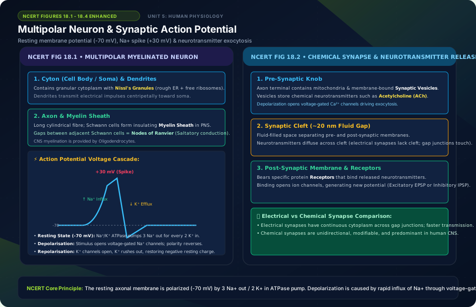
Multipolar Neuron & Synaptic Action Potential
🚨 Examiner Traps & Common Misconceptions
✅ Reality: The resting axoplasm has low Na+ and high K+! The Na+/K+ pump actively expels 3 Na+ ions out for every 2 K+ ions pumped inside.
✅ Reality: RODS contain rhodopsin for twilight (scotopic) vision. CONES provide daylight (photopic) color vision (Red, Green, Blue cones).
💡 Memory Hack & Mnemonic
Sodium-Potassium ATPase Ratio: ‘NOKIA: Na Out, K In Always’ (3 Na+ Out, 2 K+ In).
⭐ 10/10 Active Recall Self-Check: Chapter 18
Test your mastery before closing this lecture note. Can you answer these without looking back?
- What are the primary diagnostic characteristics that define Neural Control and Coordination in the NCERT syllabus?
- How do the physiological or anatomical structures covered in this chapter adapt the organism to its environment?
- What are the most common trick questions examiners create based on the exceptions in this chapter?
- Can you state the core mnemonic formula for remembering the sequential stages or components described above?
⭐ 10/10 Active Recall Self-Check: Chapter 18
Test your mastery before closing this lecture note. Can you answer these without looking back?
- What are the primary diagnostic characteristics that define Neural Control and Coordination in the NCERT syllabus?
- How do the physiological or anatomical structures covered in this chapter adapt the organism to its environment?
- What are the most common trick questions examiners create based on the exceptions in this chapter?
- Can you state the core mnemonic formula for remembering the sequential stages or components described above?