
17.1 Types of Movement: Amoeboid (Macrophage pseudopodia), Ciliary (Trachea, Oviduct) & Muscular #
Movement is one of the significant features of living beings. When movements result in a change of place or location, such voluntary movements are termed locomotion (e.g., walking, running, swimming, flying).
Cells of the human body exhibit three main types of movements:
- Amoeboid Movement: Exhibited by specialized cells like macrophages and neutrophils (leukocytes) in blood. Mediated by pseudopodia produced by streaming of protoplasm (supported by microfilament cytoskeletal elements).
- Ciliary Movement: Occurs in our internal tubular organs lined by ciliated epithelium. Coordinated ciliary beating sweeps dust particles and foreign mucus out of the respiratory tract (trachea), and moves ova through the female fallopian tubes (oviducts) toward the uterus.
- Muscular Movement: Movement of our jaws, limbs, tongue, and bodily locomotion requires the contractile properties of muscles.
17.2 Skeletal Muscle Ultrastructure: Muscle Fascicles, Sarcolemma, Sarcoplasmic Reticulum & Myofibrils #
Muscle is a specialized tissue of mesodermal origin, accounting for 40 to 50% of the total body weight of a human adult. Muscles have properties of excitability, contractility, extensibility, and elasticity.
Skeletal Muscle Ultrastructure: Each organized skeletal muscle consists of numerous muscle bundles called fascicles held together by a common collagenous connective tissue sheath called fascia. Each fascicle contains many muscle fibers:
- Each muscle fiber is lined by the plasma membrane called the sarcolemma, enclosing the sarcoplasm. A muscle fiber is a syncytium (multinucleated).
- The endoplasmic reticulum of muscle fibers, called the sarcoplasmic reticulum (SR), is the master cellular storehouse of Calcium ions (Ca2+).
- The sarcoplasm contains parallelly arranged myofibrils exhibiting alternate light and dark bands:
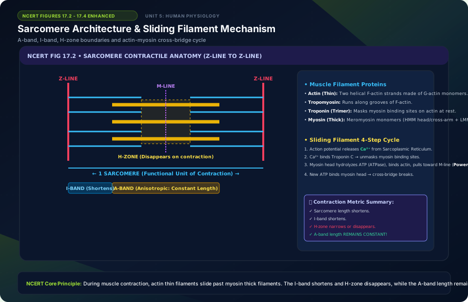
- I-Band (Isotropic / Light Band): Contains thin actin filaments; bisected by an elastic membrane called the Z-line.
- A-Band (Anisotropic / Dark Band): Contains thick myosin filaments; central lighter zone where actin does not overlap is called the H-zone, bisected by a thin fibrous M-line.
- The portion of a myofibril between two successive Z-lines is the functional contractile unit called a Sarcomere.
17.3 Contractile Proteins: Thin Actin Filaments (F-actin, Tropomyosin, Troponin) & Thick Myosin Filaments (Meromyosin, Heavy & Light chains) #
A sarcomere contains two primary contractile protein filaments:
- Thin Filament (Actin): Composed of three protein components:
- F-Actin (Filamentous Actin): Two helically wound strands of polymerized globular G-actin monomers.
- Tropomyosin: Two fibrous protein filaments that run close to the F-actin grooves along their entire length. In a resting muscle, tropomyosin covers and masks the active myosin-binding sites on actin.
- Troponin: A complex trimeric protein distributed at regular intervals on tropomyosin. Contains a subunit (Troponin C) that has a high affinity for binding Ca2+ ions.
- Thick Filament (Myosin): A polymer composed of many monomeric proteins called Meromyosins. Each meromyosin has two parts:
- Heavy Meromyosin (HMM): A globular head with a short arm projecting outwards as a cross-bridge. The globular head possesses an actin-binding site and an active ATPase enzyme site that binds and hydrolyzes ATP.
- Light Meromyosin (LMM): The linear structural tail.
17.4 Sliding Filament Theory of Muscle Contraction: Neuromuscular Junction, Acetylcholine, Ca2+ Release from SR, Cross-Bridge Cycling #
The mechanism of muscle contraction is explained by the Sliding Filament Theory, which states that contraction occurs by the sliding of thin actin filaments over thick myosin filaments:
- A motor neuron transmits an action potential arriving at the Neuromuscular Junction (Motor End Plate), releasing the neurotransmitter Acetylcholine (ACh) into the synaptic cleft.
- ACh generates an action potential across the sarcolemma, which spreads deep into the muscle fiber via T-tubules, stimulating the sarcoplasmic reticulum to release Ca2+ ions into the sarcoplasm.
- Ca2+ binds to Troponin C on the actin filament, inducing conformational changes that remove the tropomyosin mask from the active myosin-binding sites.
- The myosin globular head hydrolyzes ATP (
ATP → ADP + Pi), using the released energy to bind to the exposed active site on actin, forming a cross-bridge. - The myosin head tilts, pulling the attached actin filaments inward toward the center of the A-band (the power stroke). This pulls the Z-lines closer together, causing shortening of the sarcomere (contraction). During contraction, the I-bands shorten, the H-zone disappears, while the A-band retains its length!
- A new ATP molecule binds to the myosin head, breaking the cross-bridge. The cycle repeats as long as Ca2+ and ATP remain available. When neural signals cease, Ca2+ is pumped back into the SR, tropomyosin masks actin again, and the muscle relaxes.
17.5 Red Muscle Fibers (Aerobic, High Myoglobin, Abundant Mitochondria) vs White Muscle Fibers (Anaerobic, Fast Twitch) #
Skeletal muscles contain two distinct physiological types of muscle fibers based on myoglobin and mitochondrial content:
| Diagnostic Feature | Red Muscle Fibers (Slow-Twitch) | White Muscle Fibers (Fast-Twitch) |
|---|---|---|
| Myoglobin Content | Very high; gives muscles a deep reddish appearance. | Very low; pale or whitish in appearance. |
| Mitochondria Count | Abundant; specialized for oxidative phosphorylation. | Few; relies heavily on anaerobic glycolysis. |
| Sarcoplasmic Reticulum | Moderate amount of sarcoplasmic reticulum. | Extensive, highly developed sarcoplasmic reticulum. |
| Contraction Speed & Fatigue | Slow, sustained contraction; highly resistant to fatigue (e.g., extensor back muscles). | Fast, powerful contractions; fatigues rapidly due to lactic acid accumulation (e.g., eyeball muscles). |
17.6 Human Skeletal System: Axial Skeleton (80 bones: Skull, Vertebral Column, Ribs, Sternum) & Appendicular Skeleton (126 bones: Limbs, Pectoral & Pelvic Girdles) #
The human skeletal system consists of 206 bones and a few cartilages, divided into the Axial Skeleton and Appendicular Skeleton:
- Axial Skeleton (80 bones): Arranged along the main longitudinal body axis:
- Skull (22 bones): 8 cranial bones (Frontal, Parietal [2], Temporal [2], Occipital, Sphenoid, Ethmoid) + 14 facial bones. A single U-shaped hyoid bone is present at the base of the buccal cavity. 6 ear ossicles (Malleus, Incus, Stapes in pairs). The human skull is dicondylic (two occipital condyles articulating with the atlas vertebra).
- Vertebral Column (26 serially arranged vertebrae): 7 Cervical (C1 is Atlas, C2 is Axis), 12 Thoracic, 5 Lumbar, 1 Sacral (fused), and 1 Coccygeal (fused).
- Sternum: A flat bone on the ventral midline of the thorax.
- Ribs (12 pairs): True ribs (pairs 1–7, attach dorsally to thoracic vertebrae and ventrally to sternum via hyaline cartilage); False ribs / Vertebrochondral ribs (pairs 8, 9, 10, join the 7th rib cartilage); Floating ribs (pairs 11 and 12, not connected ventrally).
- Appendicular Skeleton (126 bones):
- Forelimb (30 bones each = 60): Humerus, Radius, Ulna, Carpals (8 wrist bones), Metacarpals (5 palm bones), Phalanges (14 digits).
- Hindlimb (30 bones each = 60): Femur (thigh bone / longest and strongest), Patella (knee cap / sesamoid bone), Tibia, Fibula, Tarsals (7 ankle bones), Metatarsals (5), Phalanges (14 digits).
- Girdles: Pectoral Girdle (Clavicle / collar bone + Scapula with glenoid cavity articulating with humerus) and Pelvic Girdle (two coxal bones fused from ilium, ischium, and pubis, with the acetabulum cavity articulating with the femur head).
17.7 Joints: Fibrous (Synarthroses), Cartilaginous (Amphiarthroses) & Synovial (Ball-and-Socket, Hinge, Pivot, Gliding, Saddle) #
Joints are points of contact between bones, or between bones and cartilages, acting as fulcrums for movement. NCERT classifies joints into three major structural categories:
- Fibrous Joints (Synarthroses): Bones are held tightly by dense fibrous connective tissue; permit zero movement (e.g., immovable sutures between flat skull bones).
- Cartilaginous Joints (Amphiarthroses): Bones are joined by cartilages; permit limited movement (e.g., joints between adjacent vertebrae in the vertebral column, pubic symphysis).
- Synovial Joints (Diarthroses): Characterized by the presence of a fluid-filled synovial cavity between the articulating surfaces of two bones, allowing extensive free movement. Types of synovial joints:
- Ball and Socket Joint: Between humerus and pectoral girdle (glenoid cavity), and between femur and acetabulum.
- Hinge Joint: Knee joint, elbow joint, and interphalangeal joints.
- Pivot Joint: Between Atlas and Axis vertebrae (enables head rotation).
- Gliding Joint: Between carpals of the wrist.
- Saddle Joint: Between carpal and metacarpal of the thumb.
17.8 Musculoskeletal Disorders: Myasthenia Gravis, Tetany (Low Ca2+), Muscular Dystrophy, Arthritis, Osteoporosis & Gout (Uric Acid Crystals) #
Musculoskeletal and joint disorders detailed in NCERT include:
- Myasthenia Gravis: An autoimmune disorder affecting the neuromuscular junction, leading to progressive fatigue, weakening, and eventual paralysis of skeletal muscles.
- Muscular Dystrophy: Progressive genetic degeneration of skeletal muscle fibers, most commonly X-linked Duchenne muscular dystrophy.
- Tetany: Rapid, sustained muscular spasms and twitches due to low calcium (Ca2+) levels in body fluids (hypocalcemia).
- Arthritis: Inflammation of joints. Includes Osteoarthritis (wear-and-tear cartilage degeneration) and Rheumatoid Arthritis (autoimmune inflammation of synovial membranes).
- Gout: Inflammation of joints caused by the abnormal accumulation and crystallization of uric acid in joint cavities.
- Osteoporosis: An age-related skeletal disorder characterized by decreased bone mass and increased fragility leading to fractures. Common cause in postmenopausal women is decreased estrogen levels.
Sarcomere Architecture & Sliding Filament Mechanism
🚨 Examiner Traps & Common Misconceptions
✅ Reality: Filament lengths DO NOT CHANGE! The thin actin filaments simply slide past the thick myosin filaments, narrowing the I-band and H-zone while the A-band length stays completely constant.
✅ Reality: Tetany is caused by HYPOCALCEMIA (low blood calcium), which destabilizes membrane voltage and causes violent involuntary muscular spasms!
💡 Memory Hack & Mnemonic
Band Changes in Sarcomere Contraction: ‘H and I shrink, A stays constant’ (HA! I shrink, A stays!).
⭐ 10/10 Active Recall Self-Check: Chapter 17
Test your mastery before closing this lecture note. Can you answer these without looking back?
- What are the primary diagnostic characteristics that define Locomotion and Movement in the NCERT syllabus?
- How do the physiological or anatomical structures covered in this chapter adapt the organism to its environment?
- What are the most common trick questions examiners create based on the exceptions in this chapter?
- Can you state the core mnemonic formula for remembering the sequential stages or components described above?
⭐ 10/10 Active Recall Self-Check: Chapter 17
Test your mastery before closing this lecture note. Can you answer these without looking back?
- What are the primary diagnostic characteristics that define Locomotion and Movement in the NCERT syllabus?
- How do the physiological or anatomical structures covered in this chapter adapt the organism to its environment?
- What are the most common trick questions examiners create based on the exceptions in this chapter?
- Can you state the core mnemonic formula for remembering the sequential stages or components described above?