
15.1 Blood Composition: Plasma Proteins (Fibrinogen, Globulins, Albumin) & Formed Elements (RBCs, WBCs, Platelets) #
Blood is a specialized fluid connective tissue consisting of a straw-colored liquid matrix called plasma (55%) and cellular components called formed elements (45%).
- Plasma: Viscous fluid comprising 90–92% water and 6–8% proteins. Major plasma proteins include:
- Fibrinogen: Essential for blood clotting and coagulation.
- Globulins: Primarily involved in defense mechanisms of the body (immunoglobulins/antibodies).
- Albumin: Maintains plasma colloidal osmotic pressure. Plasma without clotting factors is called Serum.
- Formed Elements:
- Erythrocytes (Red Blood Cells – RBCs): Most abundant cells in blood (5 to 5.5 million/mm3 in a healthy male). Formed in red bone marrow. Biconcave, enucleated (lacking a nucleus in mature mammalian RBCs) to maximize space for hemoglobin (12–16 g per 100 mL of blood). Average lifespan is 120 days, after which they are destroyed in the spleen (the ‘graveyard of RBCs’).
- Leukocytes (White Blood Cells – WBCs): Nucleated cells (6000–8000/mm3) acting as the immune defense army. Divided into:
- Granulocytes: Neutrophils (60–65%, phagocytic), Eosinophils (2–3%, resist infections and associated with allergic reactions), and Basophils (0.5–1%, secrete histamine, serotonin, heparin; involved in inflammatory responses).
- Agranulocytes: Lymphocytes (20–25%, B and T lymphocytes responsible for immune responses) and Monocytes (6–8%, phagocytic cells).
- Thrombocytes (Platelets): Cell fragments produced from giant bone marrow cells called megakaryocytes (150,000–350,000/mm3). Release platelet factors essential for blood coagulation. A reduction in platelet count leads to clotting disorders and excessive internal bleeding.
15.2 Blood Groups: ABO Antigen System, Rh Factor & Erythroblastosis Fetalis #
ABO Blood Grouping: Based on the presence or absence of two surface antigen glycoproteins on RBCs: Antigen A and Antigen B, and natural antibodies present in plasma:
| Blood Group | Antigens on RBCs | Antibodies in Plasma | Donor Compatibility |
|---|---|---|---|
| A | A | Anti-B | A, O |
| B | B | Anti-A | B, O |
| AB | Both A and B | Nil | Universal Recipient (AB) |
| O | Nil | Both Anti-A and Anti-B | Universal Donor (O) |
Rh Grouping & Erythroblastosis Fetalis: Another antigen, the Rh antigen (first discovered in Rhesus monkeys), is observed on the surface of RBCs in nearly 80% of humans (Rh positive, Rh+). If an Rh-negative mother carries an Rh-positive fetus, maternal blood can be exposed to fetal Rh antigens during delivery of the first child, developing anti-Rh antibodies. In subsequent pregnancies with an Rh+ fetus, maternal anti-Rh antibodies cross the placenta into the fetus, destroying fetal RBCs. This severe condition is called Erythroblastosis fetalis, resulting in fatal anemia and jaundice. It is prevented by administering anti-Rh antibodies (Rhogam) to the mother immediately after the first delivery.
15.3 Coagulation of Blood: Clotting Cascade, Thromboplastin, Prothrombinase, Thrombin & Fibrin Mesh #
When blood vessels are cut, bleeding stops within a few minutes due to the formation of a dark reddish-brown clot (coagulum). This process is called coagulation (blood clotting).
Coagulation is a complex enzymatic cascade involving inactive clotting factors present in plasma:
- An injured tissue and damaged platelets release thromboplastin.
- Thromboplastin helps form an active enzyme complex called thrombokinase (prothrombinase) in the presence of Calcium ions (Ca2+).
- Thrombokinase converts the inactive plasma protein prothrombin into active thrombin.
- Thrombin acts as a proteolytic enzyme to convert soluble fibrinogen into insoluble fibrin threads.
- Fibrin threads form an interlocking meshwork trapping dead and damaged formed elements of blood to seal the wound. Calcium ions (Ca2+) are indispensable for blood coagulation.
15.4 Lymph (Tissue Fluid): Composition, Functions & Lacteal Fat Absorption #
As blood passes through capillaries in tissues, water along with small water-soluble solutes filters out into intercellular spaces, leaving large proteins and formed elements in blood vessels. This filtered fluid is called interstitial fluid or tissue fluid.
Tissue fluid has the same mineral distribution as plasma and mediates the exchange of nutrients and gases between blood and cells. An elaborate network of lymphatic vessels collects this fluid and drains it back into major veins; this collected fluid is called Lymph.
Lymph is a colorless fluid containing specialized lymphocytes responsible for immune responses. Importantly, fats are absorbed through lymph in specialized lymphatic capillaries called lacteals present in the intestinal villi.
15.5 Circulatory Pathways: Open vs Closed; Single vs Incomplete vs Double Circulation #
The circulatory patterns across animal phyla are classified into:
- Open Circulatory System: Blood pumped by the heart passes through large vessels into open body cavities and spaces called sinuses, directly bathing tissues (Arthropods and Molluscs).
- Closed Circulatory System: Blood is pumped by the heart and circulates strictly within a closed network of blood vessels (Annelids and Chordates). This pattern is physiologically advantageous because fluid flow can be precisely regulated.
- Vertebrate Heart Evolutionary Progression:
- Fishes: 2-chambered heart (1 atrium, 1 ventricle) exhibiting single circulation (heart pumps deoxygenated blood to gills, which oxygenate blood and send it directly to body tissues).
- Amphibians & Reptiles (except crocodiles): 3-chambered heart (2 atria, 1 ventricle) exhibiting incomplete double circulation (oxygenated blood from lungs/skin and deoxygenated blood from body mix in the single ventricle).
- Crocodiles, Birds & Mammals: 4-chambered heart (2 atria, 2 ventricles) with completely separated pulmonary and systemic circuits: true double circulation with zero mixing of oxygenated and deoxygenated blood.
15.6 Human Heart Anatomy: 4 Chambers, Atrioventricular Valves (Tricuspid & Bicuspid/Mitral), Semilunar Valves #
The human heart is a mesodermally derived muscular organ situated in the thoracic cavity between the lungs, tilted slightly to the left, enclosed in a double-walled membranous bag called the pericardium filled with pericardial fluid.
The heart has four chambers: two upper receiving atria and two lower pumping ventricles:
- The walls of the ventricles are much thicker than the atria (the left ventricular wall is the thickest because it pumps blood against high systemic vascular resistance).
- Atrioventricular (AV) Valves: Prevent backflow of blood from ventricles into atria:
- Tricuspid Valve: Located between right atrium and right ventricle, consisting of three muscular cusps.
- Bicuspid (Mitral) Valve: Located between left atrium and left ventricle, consisting of two cusps.
- Semilunar Valves: Crescent-shaped pocket valves guarding the openings of the right ventricle into the pulmonary artery and the left ventricle into the systemic aorta, preventing regurgitation of blood back into the ventricles during diastole.
15.7 Cardiac Conduction System: SA Node (Pacemaker ~72 bpm), AV Node, Bundle of His & Purkinje Fibers #
The human heart is myogenic, meaning its normal rhythmic contractions are generated auto-rhythmically by specialized nodal musculature without requiring external nervous stimulation:
- Sino-Atrial Node (SAN): Located in the upper right corner of the right atrium. Can generate the maximum number of action potentials (70–75 action potentials/minute) and sets the basic pace of the heart. Hence, the SAN is called the Pacemaker of the heart.
- Atrio-Ventricular Node (AVN): Located in the lower left corner of the right atrium near the atrioventricular septum; relays impulses with a brief delay (0.1s) allowing atria to finish emptying into ventricles.
- AV Bundle (Bundle of His): Originates from AVN, passes through the interventricular septum, and divides into right and left bundle branches.
- Purkinje Fibers: Fine conducting fibers radiating throughout the ventricular myocardium, driving rapid, synchronized upward contraction of both ventricles.
15.8 The Cardiac Cycle: Atrial Systole, Ventricular Systole, Joint Diastole (0.8s) & Heart Sounds (LUB-DUB) #
The sequence of electrical and mechanical events occurring from the beginning of one heartbeat to the beginning of the next is called a Cardiac Cycle. At a resting rate of 72 beats/minute, the duration of one cardiac cycle is 0.8 seconds:
- Joint Diastole (0.4s): All four chambers are relaxed. Tricuspid and bicuspid valves are open; blood flows passively from vena cava and pulmonary veins into atria and on into ventricles. Semilunar valves are closed.
- Atrial Systole (0.1s): The SAN generates an action potential, stimulating simultaneous contraction of both atria. This pumps remaining blood into ventricles, increasing ventricular filling by about 30%.
- Ventricular Systole (0.3s): The action potential reaches ventricles via AVN and Purkinje fibers. Ventricular contraction increases ventricular pressure, immediately closing the tricuspid and bicuspid valves to prevent backflow (producing the first heart sound: LUB). When ventricular pressure exceeds aortic/pulmonary pressure, semilunar valves open, ejecting blood into circulation.
- Ventricular Diastole: Ventricles relax, ventricular pressure falls, causing immediate closure of the semilunar valves (producing the second heart sound: DUB).
Each ventricle pumps out approximately 70 mL of blood per cardiac cycle, which is called the Stroke Volume (SV). The Cardiac Output (CO) is the volume of blood pumped by each ventricle per minute: CO = Stroke Volume × Heart Rate = 70 mL × 72 bpm ≈ 5000 mL (5 Liters/min).
15.9 Electrocardiogram (ECG): P Wave (Atrial Depolarization), QRS Complex (Ventricular Depolarization), T Wave (Ventricular Repolarization) #
An Electrocardiograph is an instrument used to record the electrical potentials generated by the heart muscle during each cardiac cycle, producing a graphical representation called an Electrocardiogram (ECG). A standard clinical ECG is recorded with 3 electrical leads (one on each wrist and one on the left ankle):
- P-Wave: Represents the depolarization of the atria, leading to atrial contraction (systole).
- QRS Complex: Represents the depolarization of the ventricles, which initiates ventricular contraction. Ventricular systole begins shortly after the Q wave. By counting the number of QRS complexes in a given time period, a clinician determines the heart rate of an individual!
- T-Wave: Represents the repolarization of the ventricles (return from excited to normal resting state). The end of the T-wave marks the end of ventricular systole.
15.10 Disorders: Hypertension (High BP), Coronary Artery Disease (Atherosclerosis), Angina Pectoris & Heart Failure #
Circulatory system disorders detailed in NCERT include:
- Hypertension (High Blood Pressure): Normal resting blood pressure is 120/80 mmHg (120 mmHg systolic, 80 mmHg diastolic). Sustained blood pressure readings of 140/90 mmHg or higher indicate hypertension, which can damage vital organs like the brain, kidneys, and heart.
- Coronary Artery Disease (CAD) / Atherosclerosis: Deposition of calcium, fat, cholesterol, and fibrous tissue inside the coronary arteries supplying the heart muscle, narrowing the arterial lumen and restricting blood flow.
- Angina Pectoris (‘Strangling Chest Pain’): Acute chest pain that appears when insufficient oxygen reaches the heart myocardium.
- Heart Failure: The state of the heart when it cannot pump blood effectively enough to meet the metabolic needs of the body (often called congestive heart failure because lung congestion is a major symptom). Distinct from cardiac arrest (heart stops beating completely) and heart attack (heart muscle is suddenly damaged by inadequate blood supply).
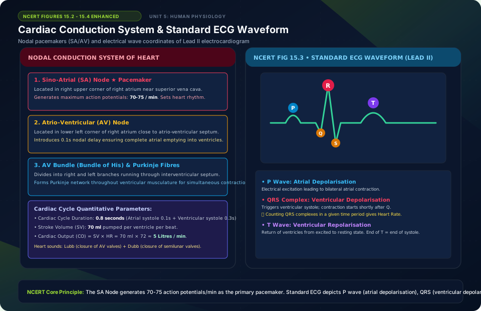
Cardiac Conduction System & Standard ECG Waveform
🚨 Examiner Traps & Common Misconceptions
✅ Reality: LUB is caused by closure of Atrioventricular (Tricuspid and Mitral) valves! DUB is caused by closure of Semilunar valves.
✅ Reality: O negative red blood cells have ZERO ABO antigens and ZERO Rh antigens, making them universal red cell donors!
💡 Memory Hack & Mnemonic
WBC Abundance in Blood: ‘Never Let Monkeys Eat Bananas’ (Neutrophils 60-65% > Lymphocytes 20-25% > Monocytes 6-8% > Eosinophils 2-3% > Basophils 0.5-1%).
⭐ 10/10 Active Recall Self-Check: Chapter 15
Test your mastery before closing this lecture note. Can you answer these without looking back?
- What are the primary diagnostic characteristics that define Body Fluids and Circulation in the NCERT syllabus?
- How do the physiological or anatomical structures covered in this chapter adapt the organism to its environment?
- What are the most common trick questions examiners create based on the exceptions in this chapter?
- Can you state the core mnemonic formula for remembering the sequential stages or components described above?
⭐ 10/10 Active Recall Self-Check: Chapter 15
Test your mastery before closing this lecture note. Can you answer these without looking back?
- What are the primary diagnostic characteristics that define Body Fluids and Circulation in the NCERT syllabus?
- How do the physiological or anatomical structures covered in this chapter adapt the organism to its environment?
- What are the most common trick questions examiners create based on the exceptions in this chapter?
- Can you state the core mnemonic formula for remembering the sequential stages or components described above?