
10.1 Cell Cycle Phases: Interphase (G1, S, G2) & M Phase; Quiescent G0 Checkpoint #
Growth and reproduction are universal characteristics of all living organisms. A cell reproduces by dividing into two daughter cells, which in turn grow and divide, forming a cellular lineage. The sequence of events by which a cell duplicates its genome, synthesizes the other constituents of the cell, and eventually divides into two daughter cells is termed the cell cycle.
Although cell growth (in terms of cytoplasmic increase) is a continuous process, DNA synthesis occurs only during one specific stage of the cell cycle. The replicated chromosomes (DNA) are then distributed to daughter nuclei through a complex series of events during cell division under strict genetic control.
A typical human cell in culture divides approximately once every 24 hours, though the cycle duration varies across organisms (e.g., yeast completes a cell cycle in only 90 minutes!). The cell cycle is divided into two basic phases:
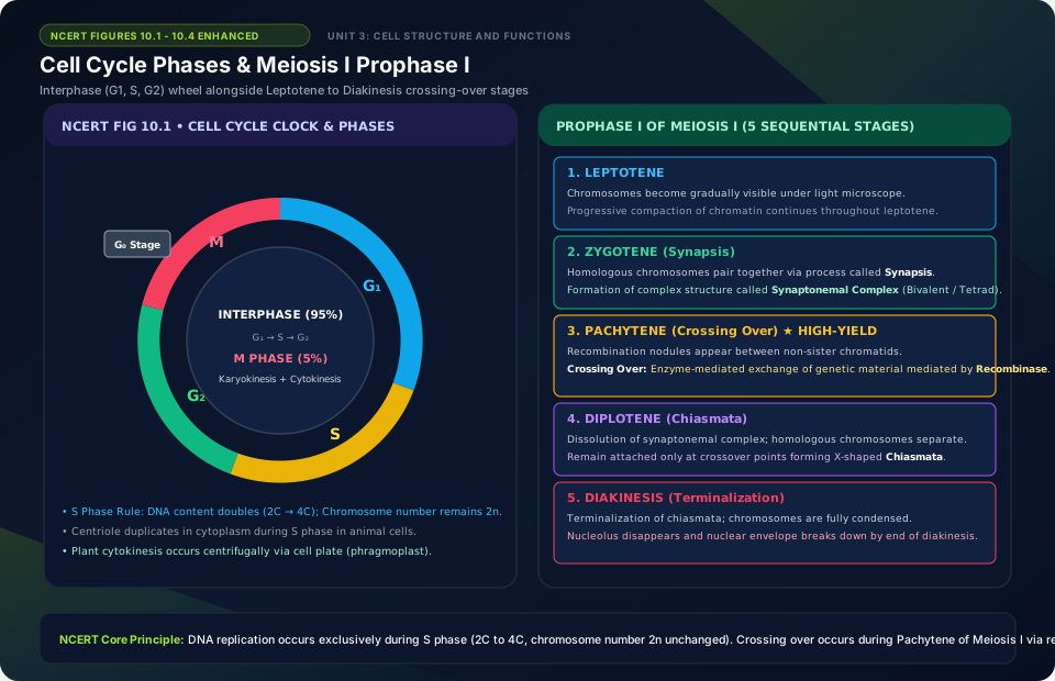
- Interphase (Resting Phase): Represents the phase between two successive M-phases, occupying more than 95% of the total duration of the cell cycle. The cell prepares for division by undergoing both cell growth and DNA replication in an orderly manner. Interphase is divided into three distinct phases:
- G1 Phase (Gap 1): The interval between mitosis and initiation of DNA replication. The cell is metabolically highly active and continuously grows, synthesizing RNA, proteins, and organelles, but does not replicate its DNA.
- S Phase (Synthesis): Marks the phase where DNA replication occurs. The amount of DNA per cell doubles (e.g., if initial amount is 2C, it increases to 4C). Crucial NCERT fact: However, there is no increase in chromosome number; if the cell had diploid (2n) chromosomes in G1, it still has 2n chromosomes after S phase! In animal cells, the centriole duplicates in the cytoplasm during S phase.
- G2 Phase (Gap 2): Proteins needed for mitosis (such as tubulin for spindle fibers) are synthesized while cell growth continues.
- G0 Phase (Quiescent Stage): Some adult animal cells do not divide (e.g., heart muscle cells, neurons), or divide only occasionally to replace injured or lost cells. These cells exit the G1 phase to enter an inactive metabolic stage called the G0 phase. Cells in G0 remain metabolically active but do not proliferate unless called upon to do so.
10.2 S Phase Molecular Events: DNA Replication & Centriole Duplication #
The M-Phase (Mitosis) is the equational division phase where the duplicated cellular components are distributed equally into two identical daughter cells. In animals, mitotic division is seen only in diploid somatic cells (with rare exceptions like male honeybees), whereas plants exhibit mitosis in both haploid and diploid cells.
Mitosis is divided into nuclear division (Karyokinesis) and cytoplasmic division (Cytokinesis). Karyokinesis progresses through four continuous, highly coordinated morphological stages:
- Prophase: Chromatin material condenses into distinct chromosomes. Centrosomes that duplicated during S phase begin moving toward opposite poles, radiating microtubular asters. The two asters together with spindle fibers form the mitotic apparatus. By the end of prophase, the nucleolus, endoplasmic reticulum, Golgi complexes, and nuclear envelope disappear.
- Metaphase: Nuclear envelope is completely broken down. Chromosomes are shortest and thickest, making metaphase the best stage to study chromosome morphology. Each chromosome consists of two sister chromatids held together at the centromere. Spindle fibers attach to the kinetochores of chromosomes. Chromosomes align along the cell’s equatorial plane, called the metaphase plate.
- Anaphase: The centromere of each chromosome splits simultaneously, and the two sister chromatids separate to become daughter chromosomes of the future daughter nuclei. As spindle fibers shorten, the daughter chromosomes migrate toward opposite poles. The centromere leads toward the pole with the chromosome arms trailing behind, displaying characteristic shapes (V for metacentric, L for sub-metacentric, J for acrocentric, I for telocentric).
- Telophase: Chromosomes reach their respective poles, decondense, and lose their individuality, reforming a diffuse chromatin mass. A new nuclear envelope assembles around each chromosome cluster. The nucleolus, Golgi complex, and ER reform. Karyokinesis is now complete.
10.3 Mitosis Stages: Prophase, Metaphase (Equatorial Plate), Anaphase (Centromere Split), Telophase & Cytokinesis #
Following the completion of nuclear division (karyokinesis), the parent cell divides its cytoplasm into two daughter cells by the process of cytokinesis. The mechanism of cytokinesis differs fundamentally between animal and plant cells due to the presence of a rigid cell wall in plants:
- Cytokinesis in Animal Cells (Cleavage Furrow Method): In animal cells, cytokinesis is achieved by the appearance of a furrow in the plasma membrane. A contractile ring of actin and myosin filaments forms beneath the membrane. The furrow gradually deepens centripetally (from the periphery toward the center) until it meets in the middle and cleaves the cell into two.
- Cytokinesis in Plant Cells (Cell Plate Method): Plant cells are enclosed by an inextensible rigid cell wall, so they cannot form a furrow. Instead, cytokinesis begins in the center of the cell and grows centrifugally (from the center outward toward the lateral walls). Precursor vesicles derived from the Golgi apparatus align at the equator to form a fragile structure called the phragmoplast (cell plate), which matures into the calcium-pectate middle lamella between the walls of adjacent daughter cells.
Significance of Syncytium: In some organisms, karyokinesis is not immediately followed by cytokinesis. Consequently, a multinucleated condition arises, termed a syncytium (e.g., the liquid endosperm in coconut).
10.4 Significance of Mitosis: Growth, Tissue Repair & Maintaining Nucleo-Cytoplasmic Ratio #
Mitosis, or equational division, is of supreme evolutionary and physiological importance to multicellular organisms:
- Conservation of Genetic Identity: Mitosis results in the production of diploid daughter cells with identical genetic complements, ensuring genetic stability across all somatic cell lineages.
- Growth and Development: The growth of multicellular organisms from a single-celled zygote into an adult consisting of trillions of cells is entirely due to mitotic divisions.
- Restoration of Nucleo-Cytoplasmic Ratio: As a cell grows, its volume increases faster than its nuclear surface area, disrupting the nucleo-cytoplasmic ratio. Mitosis divides the cell, restoring the optimal nucleo-cytoplasmic balance essential for cellular coordination.
- Cell Repair and Tissue Regeneration: Cells of the upper epidermal layer of skin, cells of the lining of the gut, and circulating blood cells are continuously lost and replaced by mitotic division. In plants, continuous vegetative growth throughout life occurs via active mitosis in apical and lateral meristems.
10.5 Meiosis I Reductional Division: Prophase I Sub-stages (Leptotene, Zygotene, Pachytene, Diplotene, Diakinesis) #
Sexual reproduction involves the fusion of two haploid gametes (each with ‘n’ chromosomes) to form a diploid zygote (‘2n’). To maintain a constant chromosome number from generation to generation in sexually reproducing species, organisms undergo a specialized reductional division called Meiosis.
Meiosis reduces the chromosome number by half: a single diploid parent cell produces four haploid daughter cells. Meiosis involves two sequential cycles of nuclear and cell division—Meiosis I and Meiosis II—but only a single cycle of DNA replication (during interphase before Meiosis I).
Meiosis I (Reductional Division): Separates homologous chromosomes, reducing chromosome number from 2n to n. Prophase I is exceptionally long and complex, divided into 5 distinct sub-stages:
- Leptotene: Chromatin condenses, and chromosomes become gradually visible under the light microscope as long, thin threads.
- Zygotene: Homologous chromosomes begin pairing together lengthwise in a process called synapsis. This pairing is mediated by the assembly of a complex proteinaceous structure called the synaptonemal complex. The paired homologous chromosomes are called a bivalent or a tetrad (four chromatids).
- Pachytene: The bivalent chromosomes appear clearly as tetrads. Crossing over occurs between non-sister chromatids of homologous chromosomes! Crossing over is the exchange of genetic material mediated by the enzyme recombinase. Recombination nodules appear where non-sister chromatids cross over, resulting in new genetic combinations.
- Diplotene: The synaptonemal complex dissolves, and the homologous chromosomes separate from each other except at the specific sites of crossover. These X-shaped points of crossing over are called chiasmata (singular: chiasma). In oocytes of some vertebrates, diplotene can last for months or years (dictyotene stage).
- Diakinesis: Final stage of Prophase I, marked by terminalization of chiasmata (chiasmata slip off to the chromosome ends like a zipper). Chromosomes are fully condensed, the meiotic spindle is assembled, and the nucleolus and nuclear envelope disappear.
10.6 Synapsis, Synaptonemal Complex, Crossing Over (Recombinase) & Chiasmata Resolution #
Following Prophase I, Meiosis I continues through:
- Metaphase I: Bivalent chromosomes align on the equatorial plate. Spindle fibers from opposite poles attach to the kinetochores of homologous chromosome pairs.
- Anaphase I: Homologous chromosomes separate and move toward opposite poles, while sister chromatids remain associated at their centromeres! (Contrast with mitosis, where centromeres split).
- Telophase I: Nuclear membrane and nucleolus reappear; the cell divides into two daughter cells (dyad of cells), each containing ‘n’ chromosomes with two chromatids.
- Interkinesis: The brief resting stage between Meiosis I and Meiosis II. Crucial fact: There is no DNA replication during interkinesis!
Meiosis II (Equational Division): Resembles a normal mitotic division, separating the two sister chromatids of each chromosome. Progresses through Prophase II, Metaphase II (chromosomes align at equator), Anaphase II (centromeres split, allowing sister chromatids to move to opposite poles), and Telophase II. Cytokinesis produces a tetrad of four genetically unique haploid daughter cells.
- Ensures conservation of the specific chromosome number of each species across generations.
- Generates novel genetic variations through crossing over and independent assortment of maternal and paternal chromosomes, providing raw material for natural selection and evolution.
10.7 Meiosis II Equational Division & Significance: Genetic Variation & Gametogenesis #
Meiosis II Equational Division & Significance: Genetic Variation & Gametogenesis is a core concept in this chapter. Understanding this requires analyzing its structural organization, biological mechanisms, and regulatory feedback pathways.
Cell Cycle Phases & Meiosis I Prophase I
🚨 Examiner Traps & Common Misconceptions
✅ Reality: In S phase, DNA content doubles (from 2C to 4C), but the chromosome number remains strictly constant (2n)!
✅ Reality: Centromeres do NOT split in Anaphase I; homologous chromosome pairs separate. Centromeres split only in Anaphase II (and mitotic anaphase).
💡 Memory Hack & Mnemonic
Prophase I Sub-stages: ‘Lazy Zebras Pack Delicious Drinks’ (Leptotene, Zygotene, Pachytene, Diplotene, Diakinesis).
⭐ 10/10 Active Recall Self-Check: Chapter 10
Test your mastery before closing this lecture note. Can you answer these without looking back?
- What are the primary diagnostic characteristics that define Cell Cycle and Cell Division in the NCERT syllabus?
- How do the physiological or anatomical structures covered in this chapter adapt the organism to its environment?
- What are the most common trick questions examiners create based on the exceptions in this chapter?
- Can you state the core mnemonic formula for remembering the sequential stages or components described above?
⭐ 10/10 Active Recall Self-Check: Chapter 10
Test your mastery before closing this lecture note. Can you answer these without looking back?
- What are the primary diagnostic characteristics that define Cell Cycle and Cell Division in the NCERT syllabus?
- How do the physiological or anatomical structures covered in this chapter adapt the organism to its environment?
- What are the most common trick questions examiners create based on the exceptions in this chapter?
- Can you state the core mnemonic formula for remembering the sequential stages or components described above?