
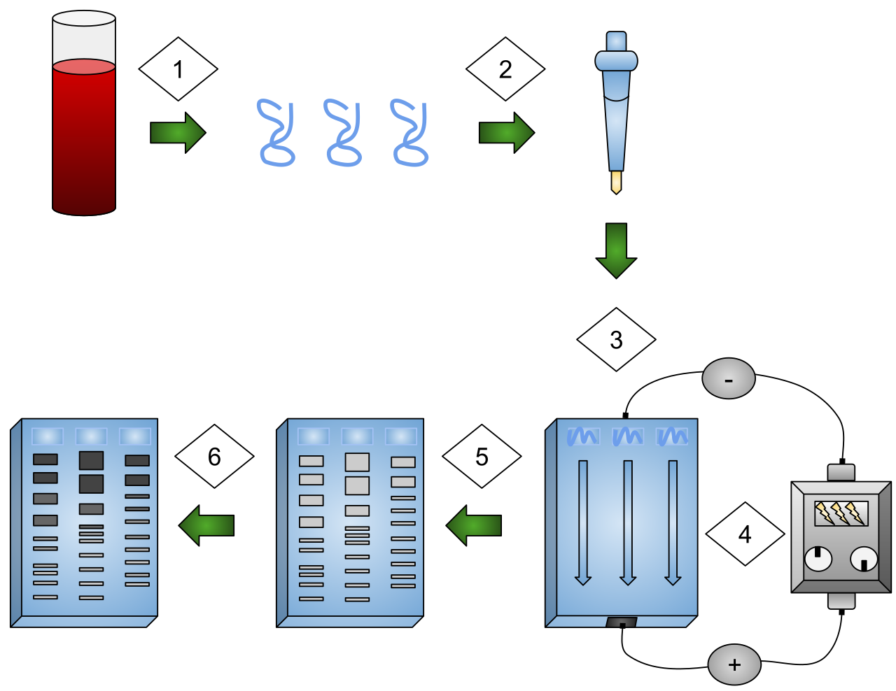
Gel Electrophoresis In Dna Fingerprinting
4.1 Selection and colour screens #
4.1.1 Antibiotic resistance is not an insert #
Selection decides who forms a colony. Screening asks which colony is right. An antibiotic plate selects the vector’s marker. Empty vector, the wrong insert, the wrong orientation, and a mutated insert all look the same.
Blue–white screening is insertional inactivation of lacZα. The fragment complements host lacZΔM15. IPTG induces; X-gal turns a working β-galactosidase colony blue. An insert in the polylinker usually makes a white colony. White means a disrupted α fragment, not a correct clone. A small in-frame insert can stay blue. A lacZα mutation with no insert, or a forgotten substrate, gives white colonies. The molecule is identified by digest and sequence.
4.2 Asking a colony a question #
4.2.1 Hybridisation and antibody screens #
Hybridisation probes DNA from colonies or plaques fixed on a membrane. Stringency sets how similar a sequence must be. A cross-species probe buys homologues and false positives. A hit can be a fragment, an intron, or a mutated copy.
An antibody screen needs an expression library. It reports an epitope, which may sit on a fragment or a cross-reacting protein, and it fails if the host never made the product or could not make the modification the antibody sees. Complementation proves an activity, not an identity, until the insert is sequenced.
4.3 Sequencing and the discipline of a map #
4.3.1 Sanger and next-generation sequencing #
Sanger sequencing stops chains with dideoxynucleotides, which lack a 3′ hydroxyl. One clean reaction reads about 500–1000 bases: junctions, a short insert, a codon. A mixed prep gives two letters at once.
Short-read sequencing, Illumina sequencing-by-synthesis being the workhorse, reads about 100–300 bases at depths that catch rare variants in a plasmid. Long reads, from Pacific Biosciences and nanopore sequencing, show rearrangements and whether the backbone is the one drawn. Junction traces do not prove an 8 kb middle. Whole-plasmid sequence is the claim “this is the construct”. Bioinformatics owns the error models.
4.3.2 Reading a plasmid map #
Trace origin, marker, and host, then promoter, ribosome-binding site, ATG, tag, stop, and terminator, in transcription order. A stop before a C-terminal tag, or a promoter pointing the wrong way, is an ordinary map error.
A claimed 6.0 kb plasmid with one BamHI site and an EcoRI site 1.5 kb away must give one 6.0 kb BamHI band, and 1.5 plus 4.5 kb from the double digest. Supercoiled uncut DNA is not a size. A third band means an extra site, star activity, or two plasmids. A missing cut suggests methylation or a missing site. Also read what the drawing implies: high copy plus a toxic gene, a lac operator titrating LacI, a mammalian polyadenylation signal that does nothing in a bacterium.
4.4 Blots and qPCR as verification #
4.4.1 What each method is allowed to say #
A Southern blot asks whether a DNA sequence is present and on what fragment. A northern blot asks the same of RNA. A western blot asks whether an antibody sees a polypeptide of the expected size. Intensity is a rough copy-number guide, not a precise one. None of the three shows a balanced pathway or a safe organism. Molecular Biology owns transfer and loading controls.
qPCR reports the cycle, Cq or Ct, at which fluorescence crosses a threshold. Earlier means more starting template only if the assay is specific and efficiencies match. The ΔΔCt model belongs to Molecular Biology. An abundant mRNA can come from a frameshifted gene that makes no protein. Proof of an expression construct is selection, a consistent digest, sequence, then a blot or an activity.
4.5 Key terms, summary, and self-check #
Key terms. Selection; screening; α-complementation; blue–white screen; hybridisation; stringency; Sanger sequencing; short-read sequencing; long-read sequencing; plasmid map; Southern blot; northern blot; western blot; Cq.
Summary. Antibiotic selection recovers a marker. Blue–white screening reports disruption of lacZα and lies in known ways. Hybridisation finds similar DNA; an antibody finds an epitope in an expression library. Sanger reads about a kilobase; short and long reads confirm a whole molecule. A map is tested by predicted fragment sizes and by tracing the expression cassette. Blots report size and presence; qPCR compares abundance under assumptions stated in Molecular Biology.
Self-check.
1. Why is every colony on ampicillin compatible with an empty vector?
2. What does a white colony report, and give two false reports?
3. A double digest yields the expected 1.5 and 4.5 kb bands plus a third. Give two explanations.
4. What do clean Sanger traces of both junctions miss in an 8 kb insert?
5. qPCR shows abundant mRNA and a western blot shows no protein. Which claim stands?
—
—
—