
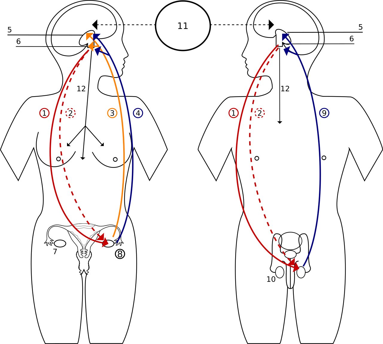
Hormons Feedback – Sprzężenie Zwrotne Hormonow
9.1 Fuel hormones #
9.1.1 Insulin #
Insulin is the hormone of the fed state. A rise in glucose is the main stimulus, sensed as in Chapter 1. Amino acids and the parasympathetic supply amplify secretion. Somatostatin and sympathetic α2 activity restrain it. The receptor is a receptor tyrosine kinase. At organ level, muscle and fat move GLUT4 to the membrane and take glucose up. The liver stops glucose output and stores glycogen. Adipose tissue stops lipolysis. Muscle takes up amino acids. The picture is storage. The intracellular pathways are Biochemistry.
Without insulin the arrows reverse. The liver produces glucose, insulin-dependent uptake fails, lipolysis supplies fatty acids, and the liver makes ketone bodies. If glucose exceeds the renal threshold of about 10–11 mmol/L, glucose in the urine obligates water, and urine volume rises. That osmotic diuresis is the renal face of a missing metabolic hormone.
9.1.2 Glucagon #
Glucagon, from alpha cells, rises when glucose is low and when amino acids are abundant, which is why a protein meal does not cause hypoglycaemia even though amino acids also release some insulin. Insulin inhibits it. The liver is the target that matters. Muscle glycogen is mobilised by contraction and by adrenaline, not by glucagon. Hepatic cAMP accelerates glycogenolysis and gluconeogenesis and, when insulin is low, ketogenesis. After a carbohydrate meal, insulin is high and glucagon low and the liver takes glucose up. Overnight, the ratio reverses and the liver holds glucose near 4–5.5 mmol/L.
9.1.3 The incretin effect #
The same glucose load releases more insulin by mouth than by vein. That difference is the incretin effect, a feed-forward signal from gut to beta cell. GIP, from upper-intestinal K cells, and GLP-1, from more distal L cells, raise beta-cell cAMP and amplify insulin secretion when glucose is already high. They do little when glucose is low, so the meal signal does not by itself drive hypoglycaemia. GLP-1 also suppresses glucagon and slows gastric emptying. The route of the glucose is part of the physiology.
9.2 Thyroid hormone and growth #
9.2.1 Synthesis and the conversion of T4 to T3 #
Follicular cells secrete thyroglobulin into the colloid. The sodium–iodide symporter concentrates iodide, and an apical exchanger, pendrin being the one usually named, moves it into the lumen. Thyroid peroxidase, using hydrogen peroxide, iodinates tyrosyl residues and couples them: two di-iodotyrosines make T4, and a mono- plus a di-iodotyrosine makes T3, still within thyroglobulin. The colloid holds weeks of hormone. TSH, through cAMP, drives uptake, synthesis, endocytosis, and gland growth. The gland secretes mostly T4.
More than 99% of circulating hormone is bound, mainly to thyroxine-binding globulin. T4’s half-life is about a week; T3’s is about a day. Deiodinases type 1 and 2 make most of the active T3 outside the thyroid. Type 3 makes inactive reverse T3. In a low-energy state, conversion shifts toward reverse T3. T3 binds a nuclear receptor and changes transcription. T3 and T4 inhibit TSH and TRH; the pituitary converts T4 to T3 locally to sense the feedback. The adult iodine requirement is small, on the order of 150 µg a day. If supply fails, hormone output falls, TSH rises, and the gland enlarges. A large iodide load transiently inhibits organification, the Wolff–Chaikoff effect, and the gland then escapes.
9.2.2 Effects on metabolic rate #
T3 raises basal metabolic rate and heat production, increasing expression of the sodium–potassium pump and of mitochondrial enzymes over hours to days. It potentiates catecholamines, partly by increasing β-receptor expression, so heart rate follows the thyroid state. Gut motility and the speed of reflex relaxation do the same. Developmental Biology owns the programme of neural development; the endocrine fact is that thyroid hormone is required while that programme runs, and a deficiency then is not fully repaired by hormone later. Too little hormone lowers heat production and slows the heart. Too much does the reverse and tends to lower weight despite appetite. TSH is the integrator most measurements use.
9.2.3 Growth hormone and IGF-1 #
Growth hormone is released in pulses, larger at night and in adolescence. GHRH stimulates, somatostatin inhibits, and ghrelin stimulates. The receptor uses JAK2–STAT, not the insulin receptor’s kinase. Linear growth is largely through IGF-1, from the liver and made locally in cartilage, acting on chondrocytes at the epiphyseal plate. IGF-1 feeds back on growth-hormone secretion. Sex steroids close the epiphyses, and after that growth hormone cannot increase height. Excess before closure lengthens bones. Excess afterwards thickens bone and soft tissue. Direct metabolic effects of growth hormone oppose insulin: lipolysis rises and muscle glucose uptake falls, while protein synthesis is supported. In fasting, IGF-1 falls and growth-hormone pulses persist, so the hormone joins glucagon, adrenaline, and cortisol in mobilising fuel.
9.3 Calcium #
9.3.1 PTH, vitamin D, and calcitonin #
About 99% of body calcium, on the order of a kilogram, is in bone. Plasma total is about 2.2–2.6 mmol/L. About half is ionised, which is the regulated and biologically active fraction; much of the rest binds albumin. A low albumin lowers total calcium without lowering the ionised fraction. Alkalosis increases albumin binding and can lower ionised calcium enough to raise neuromuscular excitability.
The calcium-sensing receptor suppresses parathyroid secretion when ionised calcium is high. A fall releases parathyroid hormone within minutes. A sustained rise increases bone resorption, so calcium and phosphate both leave bone. In the kidney, PTH increases distal calcium reabsorption, decreases proximal phosphate reabsorption, and stimulates 1α-hydroxylase. The gut effect is indirect, through calcitriol. Phosphate released from bone is excreted, so it does not precipitate with the calcium being saved.
Vitamin D3 is formed in skin from 7-dehydrocholesterol under ultraviolet light, or taken in the diet. The liver makes 25-hydroxyvitamin D, the circulating store, with a half-life of weeks. Renal 1α-hydroxylase makes calcitriol, stimulated by PTH and by low phosphate and inhibited by FGF23 and by calcitriol itself. The nuclear receptor induces intestinal calcium and phosphate absorption. In bone, calcitriol supports mineralisation by keeping the mineral supply up, and at high levels it also increases resorption.
Calcitonin, from thyroid C cells, rises when ionised calcium is high and inhibits osteoclasts. In adult humans its day-to-day role is small beside PTH and vitamin D. It matters more in some other vertebrates. FGF23, from osteocytes, is the phosphate hormone: it increases phosphate excretion and suppresses 1α-hydroxylase. PTH defends calcium and lowers phosphate. FGF23 defends phosphate.
| Hormone | Stimulus | Bone | Kidney | Gut |
|---|---|---|---|---|
| PTH | Low ionised Ca2+ | Resorption up | Ca2+ reabsorption up; phosphate reabsorption down; 1α-hydroxylase up | Indirect, via calcitriol |
| Calcitriol | PTH; low phosphate | Mineral supply supported; high levels resorb | Ca2+ and phosphate reabsorption up | Absorption of both up |
| Calcitonin | High ionised Ca2+ | Osteoclasts inhibited | Minor | Little |
| FGF23 | High phosphate | — | Phosphate excretion up; less calcitriol made | Less phosphate absorption, indirect |
9.4 The adrenal cortex and the stress response #
9.4.1 Cortisol, and the other cortical products #
The zona glomerulosa makes aldosterone under angiotensin II and potassium. The zona fasciculata makes cortisol under ACTH. The zona reticularis makes weak androgens, mainly DHEA, a significant androgen source in women and not the main male sex hormone. Cortisol feeds back on CRH and ACTH. In a person asleep at night the rhythm peaks in the hour before waking and is lowest around midnight. Stress overrides it. Steroids are made on demand; the shared slow step is mitochondrial cholesterol entry.
Cortisol raises hepatic gluconeogenesis, fills glycogen, increases muscle protein breakdown, permits lipolysis, and is permissive for catecholamine vasoconstriction. Without it, sympathetic outflow does not maintain vascular tone under stress. It also damps inflammation; the cellular detail is Immunology’s. The metabolic pattern opposes insulin and raises plasma glucose. The enzyme 11β-hydroxysteroid dehydrogenase type 2, described in Chapter 8, is what stops this larger amount of cortisol from occupying the kidney’s mineralocorticoid receptor.
9.4.2 Fight or flight #
The fast limb is sympathetic nerves plus adrenal adrenaline, in seconds. Heart rate and contractility rise (β1). Skin, gut, and kidney constrict (α1). Skeletal-muscle arterioles dilate to circulating adrenaline (β2), so output rises and is redirected. Bronchi dilate. Gut motility falls. Sweating, sympathetic but cholinergic, starts. The liver releases glucose and adipose tissue releases fatty acids, while sympathetic input suppresses the insulin that would otherwise have answered the rise in glucose. Central pathways fire this pattern from the sight or idea of a threat, before volume or glucose has changed. It is feed-forward.
Cortisol follows over minutes to hours, sustains the fuel supply, and maintains vascular responsiveness to catecholamines after the adrenaline peak has gone. Negative feedback on the ACTH axis prevents an unlimited rise. When the stress ends, insulin refills the stores. Exercise in Chapter 10 uses the same pattern for work, with one difference: working muscle locally overrides the vasoconstriction.
9.5 Key terms, summary, and self-check #
Key terms. Incretin; GIP; GLP-1; thyroglobulin; deiodinase; IGF-1; PTH; calcitriol; calcitonin; FGF23; cortisol; permissive effect; fight or flight.
Summary. Insulin stores fuels when the beta cell sees glucose, and gut incretins amplify that secretion if the glucose came by mouth. Glucagon tells the liver to release glucose. The thyroid secretes mostly T4 from iodinated thyroglobulin; deiodinases make T3, which raises metabolic rate under TSH feedback. Growth hormone, through IGF-1, drives height before the epiphyses close, and itself mobilises fat. PTH and calcitriol defend ionised calcium; FGF23 defends phosphate; calcitonin is a weak partner in adult humans. Cortisol permits catecholamines and maintains fuel supply. The fight-or-flight pattern itself is sympathetic and medullary, and it is in place within seconds.
Self-check.
1. Why does glucose by mouth release more insulin than the same glucose into a vein?
2. The thyroid secretes mostly T4. Why does T3 account for most of the effect on metabolic rate?
3. How do PTH and calcitriol divide the raising of plasma calcium among bone, kidney, and gut?
4. Why does cortisol not normally act as a mineralocorticoid on the principal cell?
5. Which parts of fight or flight are in place within seconds, and what does cortisol add later?
—
—
—