
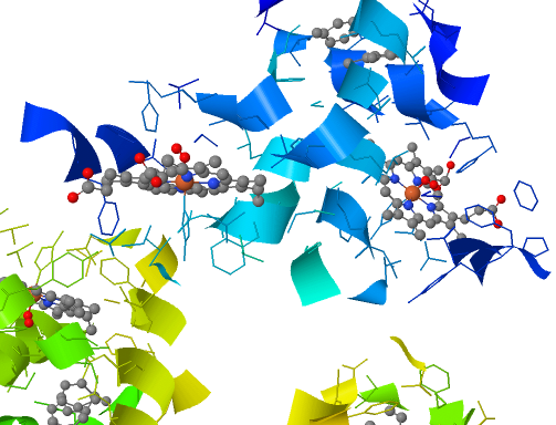
Oxyhemoglobin Active Site Structure
6.1 Plasma and cells #
6.1.1 What blood is made of #
Blood volume is about 70 mL/kg, near 5 L. A little over half is plasma. Haematocrit is about 0.40–0.50 in adult men and somewhat lower in adult women. Plasma holds sodium near 140 mmol/L, potassium near 4, chloride near 100, bicarbonate near 24, and protein near 70 g/L. Albumin, about 40 g/L, provides most of the oncotic pressure and binds poorly soluble molecules. Globulins include antibodies, which Immunology owns, and carriers. Fibrinogen is the clot’s substrate. Serum is what remains after clotting has consumed fibrinogen. Plasma and serum are not the same word.
White cells — neutrophils, eosinophils, basophils, monocytes, lymphocytes — are named only as contents. Their biology belongs to Immunology. Platelets are fragments of megakaryocytes, about 150–400 × 10^9 per litre, circulating about 8–10 days.
6.1.2 The red cell #
A mammalian red cell is a biconcave disc with no nucleus and no mitochondria. The shape aids diffusion and lets the cell deform through a narrow capillary. Birds, reptiles, amphibians, and fish have nucleated red cells; the mammalian loss of the nucleus is a specialisation, not the original vertebrate condition. Without mitochondria the cell lives on glycolysis, and 2,3-bisphosphoglycerate is a side product of that glycolysis. Haemoglobin is about 150 g/L in an adult man and somewhat lower in an adult woman. Life in the circulation is about 120 days. Production, on the order of 10^11 cells a day, is driven by erythropoietin from renal interstitial fibroblasts when local oxygen delivery falls. The count therefore rises over the weeks of altitude acclimatisation. Macrophages of spleen and liver destroy the cells. Iron is recycled. Haem becomes bilirubin, which the liver conjugates and excretes in bile.
6.2 Compatibility #
6.2.1 ABO #
Landsteiner showed that serum from one person can clump another’s red cells. The physiological fact is the mismatch between antigen on the cell and antibody in the plasma. The carbohydrate chemistry and the alleles belong to Genetics and Biochemistry.
| Group | Antigen on the red cell | Antibody in the plasma |
|---|---|---|
| O | Neither | Anti-A and anti-B |
| A | A | Anti-B |
| B | B | Anti-A |
| AB | A and B | Neither |
The antibodies are present without a previous transfusion. They are mostly IgM and are thought to follow exposure to similar carbohydrates on gut bacteria. IgM does not cross the placenta, so an ABO mismatch between mother and fetus does not usually cause the haemolytic disease below. If anti-A meets A antigen, the cells agglutinate and complement can lyse them. The recipient’s plasma must not contain antibody against the donor cells’ antigens. Group O red cells carry neither antigen, which is the limited sense in which they are general donor cells. Group O plasma contains both antibodies, so the phrase does not extend casually from red cells to whole blood.
6.2.2 Rh and haemolytic disease of the newborn #
The antigen that matters here is D. Anti-D is not naturally present in a D-negative person. It appears after exposure, and the antibody that crosses the placenta is IgG.
An Rh-negative mother carrying an Rh-positive fetus, the D inherited from the father, meets fetal red cells in small numbers during pregnancy and in larger numbers when the placenta separates. She may then make anti-D. The sensitising pregnancy is usually spared, because the response is late. In a later Rh-positive pregnancy, maternal IgG crosses the placenta and coats fetal red cells. They are haemolysed. The fetus becomes anaemic, and bilirubin from the haem accumulates. Some group-O mothers have IgG as well as IgM against A or B, so a milder version can occur with ABO incompatibility. The classical picture is Rh because anti-D is IgG and because the mother had to be primed. The mechanism is the point. Prophylaxis and treatment are not.
6.3 Haemostasis #
6.3.1 Spasm and the platelet plug #
Vascular spasm narrows a damaged small vessel within seconds, from a myogenic response, endothelin, and a sympathetic reflex. Platelets do not stick to intact endothelium, which releases prostacyclin and nitric oxide. On exposed collagen and von Willebrand factor, glycoprotein Ib binds von Willebrand factor, especially at high shear, and glycoprotein VI binds collagen. The platelet activates, makes thromboxane A2, and releases ADP. Both recruit more platelets. Glycoprotein IIb/IIIa then binds fibrinogen, which bridges platelet to platelet. The plug is local positive feedback, limited by dilution in flowing blood and by prostacyclin and nitric oxide from neighbouring intact endothelium.
6.3.2 Coagulation as a protease cascade #
Fibrin stabilises the plug. It is the end of a protease cascade that needs calcium and a phospholipid surface. A small start produces a burst of thrombin, and thrombin feeds back by activating factors V and VIII and platelets. Localisation is obligatory. Factors II, VII, IX, and X, and proteins C and S, carry γ-carboxyglutamate residues, added in the liver and dependent on vitamin K, that dock them to calcium and hence to the membrane.
| Route | Trigger | Role in a wound |
|---|---|---|
| Tissue factor | Tissue factor on cells not normally in contact with blood, plus factor VIIa, activates IX and X | The usual start |
| Contact, as in a glass tube | Factor XII, then XI, IX, VIII | Factor XII is not required for ordinary haemostasis |
| Platelet-surface amplification | The first thrombin activates platelets and factors V, VIII, and XI | Factors VIII and IX are essential; their absence bleeds |
| Common finish | Xa with Va makes thrombin; thrombin makes fibrin; XIIIa cross-links it | Shared end |
Tissue-factor pathway inhibitor soon damps the initiating complex. The burst that builds the clot is the amplification on the platelet. The test-tube story in which the contact pathway starts haemostasis in parallel describes the tube better than the wound.
6.3.3 What keeps the clot local #
Flow dilutes active factors; stasis removes that protection. Antithrombin inhibits thrombin and factor Xa, and endothelial heparan sulphate accelerates it, so inhibition is favoured on healthy wall. Thrombomodulin binds thrombin and switches it to activate protein C, which with protein S destroys factors Va and VIIIa. Thrombin on intact endothelium therefore helps stop the clot. Tissue-factor pathway inhibitor limits initiation. Prostacyclin and nitric oxide inhibit platelets. Plasmin, made from plasminogen by tissue plasminogen activator most efficiently when both are on fibrin, cuts fibrin into soluble fragments. Free plasmin and free activator are inhibited, so fibrinolysis is local too. The amplifiers are tolerable because geography and named inhibitors end them.
6.4 Key terms, summary, and self-check #
Key terms. Haematocrit; albumin; erythropoietin; ABO; Rh D; agglutination; haemolytic disease of the newborn; von Willebrand factor; thromboxane A2; tissue factor; thrombin; fibrin; antithrombin; protein C; thrombomodulin; plasmin.
Summary. Plasma carries ions, albumin, and clotting proteins. Mammalian red cells are non-nucleated bags of haemoglobin that circulate for about 120 days under erythropoietin. ABO antibodies are already present and must not meet their antigen. Anti-D appears only after exposure, is IgG, and can haemolyse the red cells of a later Rh-positive fetus. Haemostasis is spasm, a platelet plug, and fibrin. Tissue factor starts the cascade, factors VIII and IX amplify it on the platelet, and antithrombin, protein C, thrombomodulin, and plasmin keep the burst local.
Self-check.
1. Why does a person of group O already have anti-A and anti-B, and why does that not usually cause severe haemolytic disease of the newborn?
2. Why does classical Rh disease threaten a later pregnancy rather than the one in which the mother first met D?
3. What starts coagulation in a wound, and why can factor XII be absent without a bleeding illness when factor VIII cannot?
4. Name three endothelial mechanisms that oppose the clot, and say what plasmin does.
5. Why is a non-nucleated red cell a mammalian peculiarity, and about how long does it circulate?
—
—
—