
 SVG
SVGAmphibian Egg
Amphibian Egg: 1. Jelly Capsule 2. Vitelline Membrane 3. Perivitelline Fluid 4. Yolk Plug 5. Embryo The jelly capsule is designed to protect the embryo from the outside environment. This capsule also prevents the egg from drying out. The vitelline membrane is a membrane surrounding the egg. Upon fertilization, this membrane splits off from the surface of the egg and the space between the vitelline
 SVG
SVGLimb Bud
Sonic hedgehog in limb development, based on material published in Harfe BD, Scherz PJ, Nissim S, Tian H, McMahon AP and Tabin CJ (2004). "Evidence for an expansion-based temporal Shh gradient in specifying vertebrate digit identities". Cell 118 (4): 517-528.
 SVG
SVGNeural.Crest.Cells.Migration
Cross section scheme of vertebrate embryo. 1 – neural crest, 2 – neural tube, 3 – dorsal somite, 4 – notochord. Both paths of the neural crest cells migation marked by arrows: red one is the dorsolateral path, blue one signs the ventromedial path.
 SVG
SVGProtovsdeuterostomes
This diagram is showing the difference of the two major types of coelomates: the protostomes (molluscs, annelids, arthropods, ...) and deuterostomes (echinoderms, vertebrates, ...). These groups differ in several characteristics of early development; In deuterostomes blastula divisions is called "radial cleavage" because it occurs parallel or perpendicular to the major polar axis. In protostomes t
 SVG
SVGVetebrateembryo
This diagram depicts a cross section of a vertebrate embryo in the neurula stage. This stage is characterized by development of the nervous system through the formation of the neural tube. The neural tube later develops into the brain and the spinal cord. The other structures in the diagram also play major roles in development. The notochord functions as a dorsal supporting rod until it is replace
 PNG
PNGA Putative Model Of How The Organizer Induces Germ Layer Specification And Patterning
Figure 4. A putative model of how the organizer induces germ layer specification and patterning.
 PNG
PNGEarly Gastrulation In Amphibian Embryos
Figure 3. Early gastrulation in amphibian embryos. The dorsal mesoderm (organizer) cells initiate migration from the dorsal blastopore lip and establish primary germ layers. The involuting cells release several protein inhibitors that modify the surrounding cells’ fate. ECM: extracellular matrix.
 PNG
PNGEb Development And Tj Formation
Figure 2. Embryoid bodies (EB) development and tight junction (TJ) formation. (A) Schematic diagram of EB development. The development of EBs starts with the aggregation of ESCs into suspended spheroids of inner cell mass (ICM) and parallels the development of the mouse blastocyst ICM. By day 3 of culture, the outer cells differentiate into a circumferential epithelium known as the primitive endoderm (PrEn). The apical and basal domains of the PrEn face the exterior environment and interior ICM core of the EB respectively. The EBs at this point are called simple EBs. Progressively from day 4 onwards, the PrEn basally secretes key ECM components Laminins and Collagen IV to form a basement membrane (BM). The PrEn also further differentiates in the visceral endoderm (VEn). In conjunction with this, the interior ICM differentiates into the epiblast. By day 5 or 6, the epiblast in contact with the BM in turn differentiates into the primitive ectoderm (PrEc) or epiblast epithelium. The rest of the epiblast not in contact with the BM will undergo apoptosis. The apoptotic bodies are removed by autophagy-initiated phagocytosis, leaving a progressively enlarging lumen or Proamniotic-like cavity (PAC) surrounded by the apical domain of the PrEc and the basal BM. EBs at this stage are called cystic EBs and equivalent to the egg cylinder stage of the mouse peri-implantation embryo just before gastrulation. (B) Immunofluorescence staining of ZO-1 and ZO-2 in Day-5 or -6 EB cryosections. WT (panels a and e), ZO-1-/- (panels b and f), ZO-2-/- (panels c and g) and ZO-1-/- ZO-2-/- (panels d and h) EBs were stained with antibodies to ZO-1 (panels a-d, green color) or ZO-2 (panels e-h, green color). Nuclei are labeled with DAPI (blue color). Magnification of image in insets. ExEn is indicated here as ‘en’. (C) Transmission electron micrographs. TJ complex at the apico-lateral membrane of Day-9 EB ExEn was visualized by TEM as electron-dense material (indicated by arrows in magnification of inset) in WT (panels a and e), ZO-1-/- (panels b and f) and ZO-2-/- (panels c and g) EBs. This was absent in ZO-1-/- ZO-2-/- EBs (panels d and h, arrowheads in magnification of inset). (D) Immunofluorescence staining of Cldn-6 in Day-9 EB cryosections. WT, ZO-1-/-, ZO-2-/- and ZO-1-/- ZO-2-/- EBs were stained with antibodies to Cldn-6 (panels a–d, green color). Nuclei are labeled with DAPI (blue color). Magnification of image in insets. ExEn is indicated here as ‘en’.
 PNG
PNGEmbryonic Development Mouse
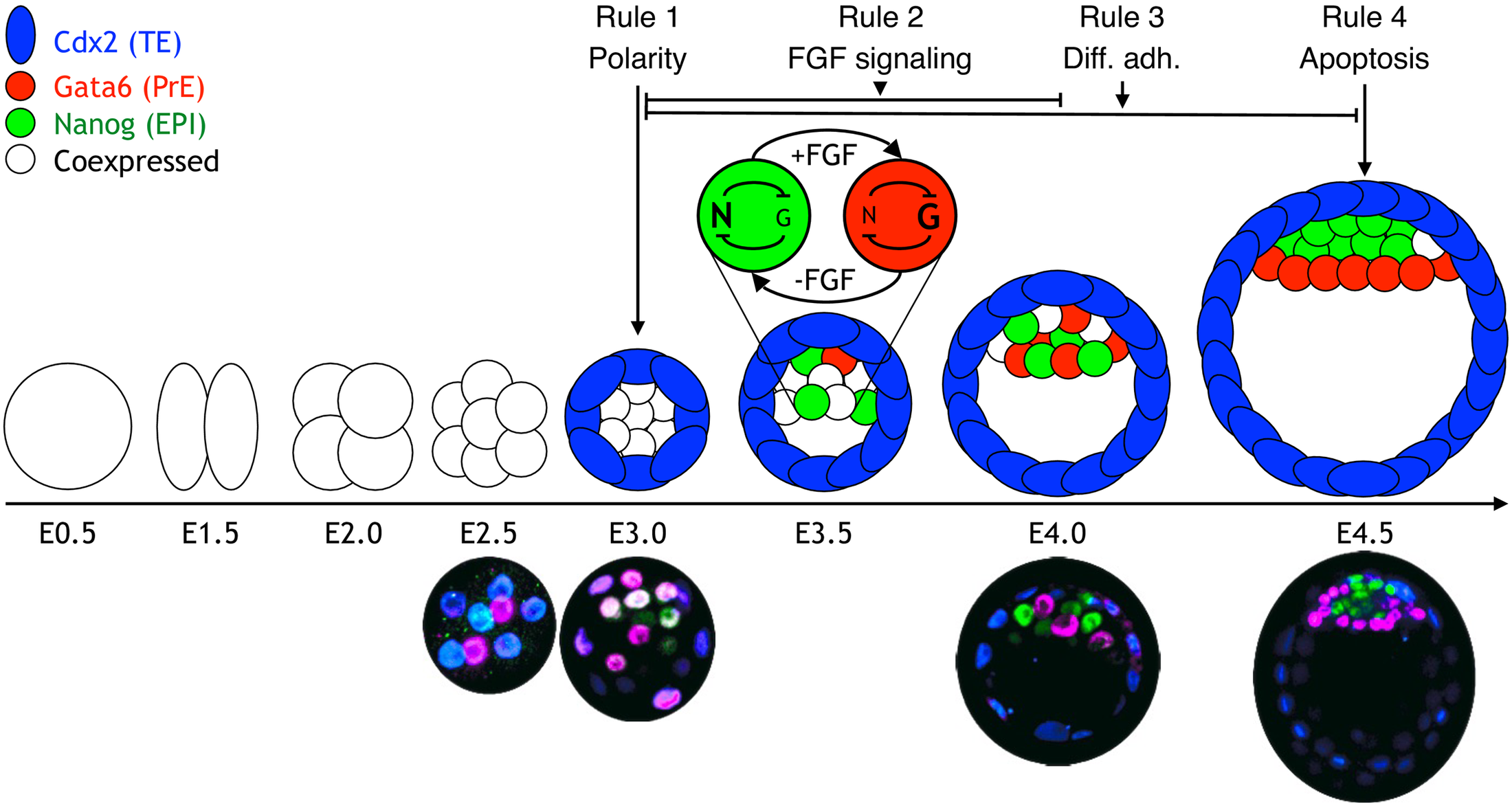
Fig 1. Schematic of the early embryonic development. The zygote (embryonic day [E] 0.5) undergoes 3 rounds of cleavage divisions, resulting in 8 cells at E2.5. During the next round of division, the blastomeres undergo compaction and become polarized, resulting in the outer trophectoderm (TE) (blue) and the inner cell mass (ICM) (still coexpressing Gata6 and Nanog). The TE expresses the transcription factor caudal-related homeobox 2 (Cdx2). At E3.5, a cavity has formed, and the ICM is positioned at 1 side of the embryo. At this stage, the ICM transcription factors, Gata6 (red) and Nanog (green), are expressed in a mutually exclusive salt-and-pepper pattern in some cells. At E4.5, Nanog- and Gata6-expressing cells have physically segregated into 2 distinct layers and are developmentally restricted to either the epiblast (EPI) or primitive endoderm (PrE). The lower panel shows immunostaining of embryos at different stages during preimplantation development. Color coding is the same as in the panel above. The timing of the 4 different rules that we apply is indicated on top of the diagram.