
5.1 The DNA Molecule: Polynucleotide Chain Architecture, Chargaff’s Equimolar Rules & Watson-Crick B-DNA Double Helix #
Deoxyribonucleic acid (DNA) and ribonucleic acid (RNA) are the two types of nucleic acids found in living systems. DNA acts as the genetic material in most organisms, while RNA acts as genetic material in some viruses (e.g., Tobacco Mosaic Virus, HIV, Poliovirus) and functions dynamically as an adapter, structural molecule, and enzyme (ribozyme).
The definitive proof that DNA is the genetic material was established through three classic experiments detailed in NCERT:
- Frederick Griffith (1928) – Transforming Principle: Griffith conducted experiments with Streptococcus pneumoniae (bacterium causing pneumonia):
- Smooth (S) strain has a mucous (polysaccharide) coat, is virulent and kills mice.
- Rough (R) strain lacks the coat, is non-virulent and mice survive.
- Heat-killed S strain injected into mice → mice survive.
- Heat-killed S strain + live R strain injected into mice → mice died! Griffith recovered living S bacteria from the dead mice. He concluded that the R strain had been transformed into virulent S strain by a heat-stable chemical compound transferred from the heat-killed S strain: the Transforming Principle.
- Oswald Avery, Colin MacLeod, and Maclyn McCarty (1944): Purified biochemicals (proteins, RNA, DNA) from heat-killed S cells. They discovered that:
- Proteases and RNases did not affect transformation.
- Treatment with DNase completely destroyed transforming activity! They proved that DNA is the transforming substance.
- Alfred Hershey and Martha Chase (1952) – The Blender Experiment: Unequivocal proof that DNA is the genetic material. They grew bacteriophage T2 on two radioactive media:
- One with radioactive phosphorus (32P), which labeled viral DNA (since DNA contains phosphorus but not sulfur).
- One with radioactive sulfur (35S), which labeled viral protein coats (since proteins contain sulfur but not phosphorus).
- After allowing viruses to infect E. coli bacteria, the culture was blended to detach viral coats and centrifuged. Only bacteria infected with 32P showed intracellular radioactivity; viral coats in supernatant had 35S. This conclusively proved that DNA is the genetic material passed from virus to bacterium!
5.2 Packaging of DNA in Eukaryotes: Histone Octamer, Nucleosome Core, Euchromatin vs Heterochromatin #
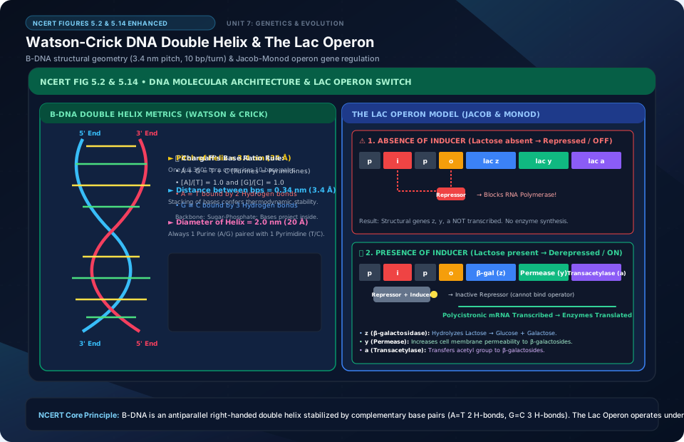
A polynucleotide chain consists of nucleotides joined by 3’–5′ phosphodiester bonds. Erwin Chargaff (1950) formulated landmark base-pairing rules for double-stranded DNA:
- The amount of Adenine is always equal to Thymine (A = T), and Guanine equals Cytosine (G = C).
- Total purines equal total pyrimidines: A + G = C + T (or
[A + G] / [C + T] = 1.0). - The base ratio
(A + T) / (G + C)is variable across different species, but constant for a given species.
In 1953, based on X-ray diffraction data produced by Maurice Wilkins and Rosalind Franklin, James Watson and Francis Crick proposed the iconic Double Helix Model of B-DNA:
- DNA is made of two polynucleotide chains where the backbone is formed of alternating sugar-phosphate groups and nitrogenous bases project inside.
- The two strands run in antiparallel polarity: one strand is 5’→3′, the other 3’→5′.
- Bases in opposite strands are paired through hydrogen bonds: A pairs with T by 2 hydrogen bonds (A = T), and G pairs with C by 3 hydrogen bonds (G ≡ C).
- The two strands are coiled in a right-handed fashion. The pitch of the helix is 3.4 nm (34 Å), with roughly 10 base pairs per turn, meaning distance between adjacent base pairs is 0.34 nm (3.4 Å).
- The plane of one base pair stacks over the other in double helix, conferring thermal and chemical stability over RNA.
5.3 The Search for Genetic Material: Griffith’s Bacterial Transformation (1928), Avery-MacLeod-McCarty Biochemical Characterization & Hershey-Chase Blender Experiment (1952 using 32P and 35S) #
The length of DNA in a typical diploid human cell is approximately 2.2 meters (6.6 × 109 bp × 0.34 × 10−9 m/bp ≈ 2.2 m). Fitting this immense 2.2-meter thread into a microscopic nucleus (~10−6 m diameter) requires extraordinary packaging:
In Eukaryotes: DNA packaging is mediated by basic, positively charged proteins called Histones. Histones are rich in the basic amino acid residues Lysine and Arginine, which carry positive charges on their side chains:
- Eight histone molecules (two copies each of H2A, H2B, H3, and H4) associate to form a Histone Octamer.
- The negatively charged DNA wraps around the positively charged histone octamer to form a structural unit called a Nucleosome. A typical nucleosome contains 200 base pairs of DNA helix.
- Nucleosomes form repeating units seen as ‘beads-on-a-string’ under an electron microscope, linked by an H1 linker histone.
- Chromatin fibers condense further with Non-Histone Chromosomal (NHC) proteins. Chromatin is organized into two distinct regions:
- Euchromatin: Loosely packed, lightly staining chromatin; transcriptionally active.
- Heterochromatin: Densely packed, intensely staining chromatin; transcriptionally inactive.
5.4 RNA World Hypothesis & Types of RNA (mRNA, tRNA ‘Cloverleaf’ Adapter, rRNA) #
Watson and Crick famously postulated: “It has not escaped our notice that the specific pairing we have postulated immediately suggests a possible copying mechanism for the genetic material.” They proposed that DNA replication is Semiconservative—the two parental strands separate, and each acts as a template for synthesizing a complementary daughter strand.
Meselson and Stahl Experiment (1958): Proved semiconservative replication in E. coli:
- Grew E. coli in medium containing heavy isotope of nitrogen (15NH4Cl) for many generations until 15N was incorporated into both strands of DNA (heavy DNA).
- Transferred bacteria into normal medium with light nitrogen (14NH4Cl). DNA was extracted and centrifuged in a Cesium Chloride (CsCl) density gradient:
- After 20 minutes (Generation 1, one cell division): DNA showed a single band at intermediate hybrid density (15N-14N)!
- After 40 minutes (Generation 2, two divisions): DNA resolved into two equal bands: 50% hybrid (15N-14N) and 50% light (14N-14N).
This conclusively proved that DNA replication is semiconservative. In a related experiment, Herbert Taylor (1958) used radioactive thymidine in broad bean (Vicia faba) root tip chromosomes to prove that DNA in chromosomes also replicates semiconservatively.
5.5 Semiconservative DNA Replication: Meselson & Stahl Experiment (15N / 14N CsCl Gradient), Replication Fork & DNA Polymerase Fidelity #
DNA replication is a high-fidelity enzymatic process catalyzed primarily by DNA-dependent DNA Polymerase:
- Replication begins at specific genomic sites called the Origin of Replication (ori). Because unwinding the entire DNA molecule requires massive energy, replication occurs inside a localized Y-shaped opening called the Replication Fork.
- DNA Polymerase III can catalyze polymerization only in the 5′ → 3′ direction. Consequently, replication is asymmetric at the replication fork:
- Leading Strand: On the 3′ → 5′ template strand, DNA synthesis proceeds continuously toward the replication fork.
- Lagging Strand: On the 5′ → 3′ template strand, DNA is synthesized discontinuously away from the fork as short fragments called Okazaki fragments.
- DNA synthesis requires an RNA primer synthesized by primase to initiate chain elongation. The RNA primers are removed and replaced with DNA, and the discontinuous Okazaki fragments are joined together by the enzyme DNA Ligase.
5.6 Transcription: Transcription Unit (Promoter, Structural Gene, Terminator), RNA Polymerases I, II, III in Eukaryotes & Post-Transcriptional Modifications (Capping, Tailing, Splicing) #
The process of copying genetic information from one strand of DNA into RNA is termed Transcription. The principle of complementarity governs transcription, with the exception that Adenine pairs with Uracil (U) instead of Thymine.
A Transcription Unit in DNA consists of three functional regions:
- A Promoter: Located towards the 5′-end (upstream) of the structural gene. It is a DNA sequence that provides the binding site for RNA Polymerase.
- The Structural Gene: The DNA segment being transcribed. The strand with 3′ → 5′ polarity acts as the Template Strand. The other strand with 5′ → 3′ polarity is called the Coding Strand (all reference points—upstream, downstream—are defined relative to the coding strand!).
- A Terminator: Located towards the 3′-end (downstream) of the coding strand, signaling the end of transcription.
In bacteria, a single DNA-dependent RNA polymerase catalyzes transcription of all three types of RNA (mRNA, tRNA, rRNA). It associates transiently with an initiation factor (sigma, σ factor) to initiate transcription at the promoter, and with a termination factor (rho, ρ factor) to terminate transcription.
5.7 The Genetic Code: George Gamow’s Triplet Code, 64 Codons, Degeneracy, Unambiguous & Universal Nature (AUG = Methionine / Start Codon) #
In eukaryotes, transcription exhibits two additional complexities:
- Three Distinct RNA Polymerases:
- RNA Polymerase I: Transcribes 28S, 18S, and 5.8S rRNAs.
- RNA Polymerase II: Transcribes precursor mRNA, called heterogeneous nuclear RNA (hnRNA).
- RNA Polymerase III: Transcribes tRNA, 5S rRNA, and snRNAs (small nuclear RNAs).
- Post-Transcriptional Modifications of hnRNA: Eukaryotic structural genes are split genes, containing coding sequences called exons interrupted by non-coding intervening sequences called introns. hnRNA undergoes three processing steps:
- Splicing: Introns are cut out and removed by a multi-protein RNA complex called the spliceosome, and exons are joined together in a defined order.
- Capping: An unusual nucleotide—7-methylguanosine triphosphate—is added to the 5′-end of hnRNA.
- Tailing (Polyadenylation): A poly-A tail containing 200 to 300 adenylate residues is added to the 3′-end in a template-independent manner. Fully processed hnRNA is now mature mRNA, which exits the nucleus into the cytoplasm for translation.
5.8 Translation: Charging of tRNA (Aminoacylation), Ribosomal Initiation, Elongation & Termination (UAA, UAG, UGA) #
The Genetic Code directs the translation of nucleotide sequences in mRNA into amino acid sequences in proteins. Physicist George Gamow argued that since there are only 4 bases (A, U, G, C) and 20 amino acids, the code must be a combination of bases: a triplet code yielding 43 = 64 codons.
Har Gobind Khorana synthesized RNA molecules with defined combinations of bases, Marshall Nirenberg developed a cell-free system for protein synthesis, and Severo Ochoa discovered polynucleotide phosphorylase to synthesize RNA with defined sequences. The genetic code dictionary possesses six salient features:
- Triplet Nature: 61 codons code for amino acids; 3 codons (UAA, UAG, UGA) do not code for any amino acid and act as Stop (Nonsense / Termination) Codons.
- Unambiguous and Specific: One codon codes for only one particular amino acid.
- Degenerate: Some amino acids are coded by more than one codon (e.g., Leucine, Serine, Arginine).
- Universal: From bacteria to humans, a codon codes for the exact same amino acid (e.g., UUU codes for Phenylalanine in all organisms). Exceptions occur in mitochondrial codons and some protozoans.
- No Punctuation: The codon is read in mRNA in a continuous fashion without punctuation.
- Dual Function of AUG: AUG codes for Methionine (Met), and acts as the universal Initiator (Start) Codon.
tRNA (The Adapter Molecule): Francis Crick postulated an adapter molecule that reads the code and binds specific amino acids. Transfer RNA (tRNA) has a clover-leaf secondary structure and an inverted L-shaped 3D structure. It has an anticodon loop with bases complementary to the mRNA codon, and an amino acid acceptor end (3′-CCA) that binds specific amino acids.
5.9 Regulation of Gene Expression in Prokaryotes: The Lac Operon Model of Jacob & Monod (Polycistronic, Inducer Lactose, Repressor Protein) #
Translation is the process of polymerizing amino acids into a polypeptide chain directed by the sequence of codons in mRNA:
- Aminoacylation of tRNA (Charging): Amino acids are activated in the presence of ATP and linked to their cognate tRNA by the enzyme aminoacyl-tRNA synthetase.
- Initiation: The small ribosomal subunit (40S in eukaryotes, 30S in prokaryotes) binds to the 5′-end of mRNA at the start codon (AUG). The initiator tRNA carrying methionine binds to AUG. The large ribosomal subunit joins, assembling the functional ribosome with two binding sites: the Peptidyl (P) site and Aminoacyl (A) site.
- Elongation: The next charged tRNA enters the A-site. A peptide bond is formed between the amino acids, catalyzed by the peptidyl transferase ribozyme (23S rRNA in bacteria). The ribosome moves codon-by-codon along the mRNA in the 5’→3′ direction (translocation).
- Termination: When a stop codon (UAA, UAG, or UGA) arrives at the A-site, a Release Factor binds to the stop codon, terminating translation and releasing the complete polypeptide.
5.10 The Human Genome Project (HGP Goals, Methodologies, Major Findings) & DNA Fingerprinting (Alec Jeffreys, VNTRs, Southern Blotting) #
Regulation of Gene Expression & The Lac Operon: In 1961, François Jacob and Jacques Monod elucidated the first genetic regulatory mechanism in E. coli: the Lac Operon (Lactose Operon), an inducible catabolic operon controlling lactose metabolism:
- The Lac Operon consists of:
- Regulator gene (i gene): Codes for the Lac Repressor protein.
- Promoter (P): Binding site for RNA polymerase.
- Operator (O): Switch site where the repressor protein binds.
- Three Structural Genes:
- z gene: Codes for β-galactosidase (hydrolyzes lactose into glucose and galactose).
- y gene: Codes for permease (increases membrane permeability to β-galactosides).
- a gene: Codes for transacetylase.
- In the Absence of Inducer (Lactose): The repressor protein binds to the operator region, preventing RNA polymerase from transcribing the operon: Switch is OFF.
- In the Presence of Inducer (Lactose / Allolactose): Lactose binds to the repressor protein, inactivating it by conformational change. The repressor cannot bind to the operator; RNA polymerase binds to the promoter and transcribes the structural genes: Switch is ON.
Human Genome Project (HGP): A 13-year mega project coordinated by the US Department of Energy and NIH, completed in 2003. Key findings: Human genome contains 3.1647 billion base pairs; average gene consists of 3000 bases (largest human gene is dystrophin with 2.4 million bases); total number of genes is estimated at ~30,000; less than 2% of the genome codes for proteins; 99.9% of nucleotide bases are exactly the same in all humans; chromosome 1 has the most genes (2968), and the Y chromosome has the fewest (231).
DNA Fingerprinting: Developed by Alec Jeffreys (1984). Based on identifying differences in specific repetitive non-coding DNA regions called Variable Number of Tandem Repeats (VNTRs) or Minisatellites (Satellite DNA) exhibiting high degrees of individual polymorphism. Involves DNA isolation, restriction digestion, agarose gel electrophoresis, Southern blotting to a nylon membrane, hybridization with radioactive VNTR probes, and detection by autoradiography.
Watson-Crick DNA Double Helix & The Lac Operon
🚨 Examiner Traps & Common Misconceptions
✅ Reality: 32P labels nucleic acids (DNA contains phosphorus, no sulfur); 35S labels proteins (methionine/cysteine contain sulfur, no phosphorus).
✅ Reality: There are 64 CODONS, but only 20 amino acids! 61 codons code for amino acids (degeneracy), and 3 codons (UAA, UAG, UGA) are non-coding stop signals.
💡 Memory Hack & Mnemonic
Three Stop Codons: ‘U Are Away, U Are Gone, U Go Away’ (UAA, UAG, UGA).
⭐ 10/10 Active Recall Self-Check: Chapter 5
Test your mastery before closing this lecture note. Can you answer these without looking back?
- What are the core experimental proofs or physiological steps that define Molecular Basis of Inheritance in the NCERT syllabus?
- How does the molecular mechanism or ecological interaction covered here operate under changing conditions?
- What are the high-frequency examiner traps or tricky numerical ratios associated with this chapter?
- Can you formulate the primary mnemonic or memory framework for remembering the key components above?
⭐ 10/10 Active Recall Self-Check: Chapter 5
Test your mastery before closing this lecture note. Can you answer these without looking back?
- What are the core experimental proofs or physiological steps that define Molecular Basis of Inheritance in the NCERT syllabus?
- How does the molecular mechanism or ecological interaction covered here operate under changing conditions?
- What are the high-frequency examiner traps or tricky numerical ratios associated with this chapter?
- Can you formulate the primary mnemonic or memory framework for remembering the key components above?