
2.1 Male Reproductive System: Testes, Seminiferous Tubules, Sertoli Cells, Leydig Cells & Accessory Ducts #
Humans are sexually reproducing and strictly viviparous organisms. The reproductive events in humans include gametogenesis (formation of gametes), insemination (transfer of sperms into female genital tract), fertilization (fusion of gametes), cleavage, blastocyst formation and implantation, gestation (embryonic development ~9 months), and parturition (childbirth).
The Male Reproductive System: Located in the pelvic region, consisting of a pair of testes, accessory ducts, accessory glands, and external genitalia:
- Testes & Scrotum: The paired oval testes are situated outside the abdominal cavity within a pouch of pigmented skin called the scrotum. The scrotum maintains the scrotal temperature at 2 to 2.5°C lower than normal internal body temperature, an absolute physiological prerequisite for normal spermatogenesis.
- Seminiferous Tubules: Each adult testis is covered by a dense fibrous capsule (tunica albuginea) and divided into about 250 testicular lobules. Each lobule contains 1 to 3 highly coiled seminiferous tubules in which sperms are produced. Each tubule is lined internally by two types of cells:
- Male Germ Cells (Spermatogonia): Diploid stem cells that undergo meiotic divisions to produce mature spermatozoa.
- Sertoli Cells (Nurse Cells): Tall columnar cells that provide mechanical support and metabolic nutrition to developing germ cells, and secrete androgen-binding protein (ABP) and inhibin.
- Interstitial / Leydig Cells: Located in the interstitial spaces outside the seminiferous tubules. Leydig cells synthesize and secrete steroid testicular hormones called Androgens (principally Testosterone) under the control of LH.
- Male Accessory Ducts: Seminiferous tubules open into the Rete testis → 10–12 fine Vasa efferentia → tightly coiled Epididymis (stores and matures sperms) → ascends as Vas deferens looping over the urinary bladder, joined by a duct from the seminal vesicle to form the Ejaculatory Duct opening into the urethra.
2.2 Female Reproductive System: Ovaries, Fallopian Tubes (Infundibulum, Ampulla, Isthmus), Uterus & Mammary Glands #
The male accessory glands comprise paired Seminal Vesicles, a single Prostate Gland, and paired Bulbourethral (Cowper’s) Glands:
- Their secretions constitute the Seminal Plasma, which is rich in fructose (the exclusive metabolic energy fuel for sperm motility), calcium ions (Ca2+), and specific clotting enzymes.
- Secretions of the bulbourethral glands are alkaline and rich in mucus, neutralizing residual acidic urine in the urethra and assisting in the lubrication of the penis prior to coitus.
- Seminal plasma combined with spermatozoa constitutes Semen. A single human ejaculate (~2.5–5 mL) contains roughly 200 to 300 million sperms. For normal fertility, at least 60% of sperms must have normal shape and size, and at least 40% must show vigorous motility.
2.3 Gametogenesis Comparison: Spermatogenesis (Spermiogenesis & Spermiation) vs Oogenesis (Unequal Cleavage & Polar Bodies) #
The Female Reproductive System: Located in the pelvic region, consisting of a pair of ovaries, oviducts, uterus, cervix, vagina, external genitalia, and a pair of mammary glands:
- Ovaries: The primary female sex organs producing the female gamete (ovum) and steroid ovarian hormones (estrogen and progesterone). Each ovary is 2–4 cm long, connected to the pelvic wall and uterus by ligaments, and enclosed by a thin epithelium covering the ovarian stroma (peripheral cortex and inner medulla).
- Fallopian Tubes (Oviducts, 10–12 cm long): Consists of three parts:
- Infundibulum: Funnel-shaped opening closest to ovary; its edges possess finger-like projections called fimbriae that collect the ovum after ovulation.
- Ampulla: The wider, curved middle part where fertilization normally takes place (ampullary region).
- Isthmus: Narrow terminal section with a thick lumen joining the uterus.
- Uterus (Womb): A single, inverted pear-shaped muscular organ supported by ligaments. The uterine wall has three tissue layers:
- Perimetrium: External thin membranous serosa.
- Myometrium: Thick middle layer of smooth muscle that exhibits strong contractions during parturition (labor).
- Endometrium: Inner glandular mucosal layer that undergoes cyclic cyclical breakdown and repair during the menstrual cycle and hosts embryo implantation.
- Mammary Glands: Paired glandular structures containing glandular tissue and variable adipose fat. The glandular tissue is divided into 15–20 mammary lobes containing clusters of cells called alveoli that secrete milk. Alveoli open into mammary tubules → mammary ducts → expanded mammary ampulla → connected to lactiferous ducts through which milk is sucked out by the infant.
2.4 The Menstrual Cycle: Follicular / Proliferative Phase, LH Surge (Day 14 Ovulation) & Luteal / Secretory Phase (Corpus Luteum & Progesterone) #
Gametogenesis is the process of gamete formation: Spermatogenesis in males and Oogenesis in females. While sharing meiotic principles, their timing and cellular dynamics differ fundamentally:
- Spermatogenesis: Begins at puberty and continues throughout adult life:
- Spermatogonia (2n = 46) on the inner tubule wall multiply mitotically.
- Some spermatogonia grow into Primary Spermatocytes (2n = 46).
- Primary spermatocyte undergoes Meiosis I to form two equal haploid Secondary Spermatocytes (n = 23).
- Secondary spermatocytes undergo Meiosis II to produce four equal haploid Spermatids (n = 23).
- Spermatids are transformed into mature spermatozoa (sperms) by the process of Spermiogenesis. After spermiogenesis, sperm heads become embedded in Sertoli cells and are finally released from seminiferous tubules into the lumen by the process of Spermiation.
- Oogenesis: Initiated during embryonic fetal development, long before birth!
- Millions of egg mother cells (oogonia, 2n) are formed within fetal ovaries; no more oogonia are formed or added after birth!
- Oogonia start meiotic division and become arrested at Prophase I of Meiosis I; these are Primary Oocytes. Each primary oocyte gets surrounded by granulosa cells, forming a primary follicle. Large numbers of follicles degenerate from birth to puberty (follicular atresia); only 60,000–80,000 primary follicles remain in each ovary at puberty.
- At puberty, under FSH stimulation, a primary follicle matures into a secondary and then a tertiary follicle (characterized by a fluid-filled cavity called the antrum).
- The primary oocyte within the tertiary follicle completes Meiosis I just prior to ovulation, producing an unequal division: a large haploid Secondary Oocyte (n = 23) and a tiny First Polar Body.
- The secondary oocyte is arrested at Metaphase II of Meiosis II! It is released from the mature Graafian follicle during ovulation. Meiosis II is completed ONLY upon the entry of a sperm into the ovum!
2.5 Fertilization at the Ampulla, Cortical Reaction (Block to Polyspermy) & Cleavage to Blastocyst #
A mature human sperm is a microscopic, flagellated motile gamete covered entirely by a plasma membrane. It measures roughly 60 µm in length, divided into four distinct regions:
- Head: Contains an elongated haploid nucleus with highly condensed chromatin. The anterior two-thirds of the head is capped by a cap-like lysosomal vesicle called the Acrosome (derived from the Golgi apparatus). The acrosome is filled with specialized proteolytic enzymes (sperm lysins: hyaluronidase and acrosin) that digest the corona radiata and zona pellucida of the ovum to facilitate fertilization.
- Neck: Short junctional region containing a proximal centriole (initiates the first cleavage division of the fertilized zygote) and a distal centriole giving rise to the axial filament.
- Middle Piece: Packed with numerous mitochondria spirally coiled around the axial filament (Nebenkern). Mitochondria generate ATP energy for vigorous flagellar tail wave motility essential for the sperm to swim through the female cervix and uterus into the fallopian tube.
- Tail: An elongated flagellum with an axonemal microtubular core extending through a sheath of cytoplasm, providing hydrodynamic propulsion.
2.6 Implantation, Placenta Function (hCG, hPL, Relaxin) & Embryonic Germ Layer Differentiation #
The reproductive cycle in female primates (monkeys, apes, and human beings) is called the Menstrual Cycle. The first menstruation begins at puberty and is called menarche. The cycle repeats at an average interval of about 28 to 29 days, terminating around age 50 as menopause.
The menstrual cycle is orchestrated by pituitary gonadotropins (FSH and LH) and ovarian hormones (Estrogen and Progesterone), divided into four chronological phases:
| Phase & Duration | Hormonal Profiles | Uterine & Ovarian Events |
|---|---|---|
| 1. Menstrual Phase (Days 1–5) | Progesterone and estrogen levels drop sharply due to degeneration of corpus luteum. | Endometrial lining breaks down; blood, tissue fluid, and unfertilized ovum are discharged as menstrual flow (40–80 mL) through the vagina. |
| 2. Follicular / Proliferative Phase (Days 6–13) | FSH secretion rises, stimulating follicle growth; growing follicles secrete Estrogen. | Primary follicle grows into mature Graafian follicle. Rising estrogen stimulates rapid regeneration and thickening of the uterine endometrium by cell proliferation. |
| 3. Ovulatory Phase (Day 14) | Both LH and FSH attain a peak level in the middle of cycle. Rapid surge in LH is called the LH Surge. | LH surge induces rupture of the mature Graafian follicle, releasing the secondary oocyte into the pelvic cavity: Ovulation. |
| 4. Luteal / Secretory Phase (Days 15–28) | The ruptured Graafian follicle transforms into a yellow glandular body called the Corpus Luteum, which secretes massive amounts of Progesterone. | Progesterone maintains and vascularizes the thick endometrium for blastocyst implantation. If fertilization does not occur, corpus luteum degenerates into a white scar (corpus albicans), progesterone drops, and a new menstrual cycle begins. |
2.7 Parturition (Foetal Ejection Reflex, Oxytocin Positive Feedback) & Lactation (Prolactin & Colostrum IgA) #
During coitus, semen is deposited by the penis into the vagina (insemination). Motile sperms swim rapidly through the cervix and uterus into the ampullary region of the fallopian tube, where the ovum released from the ovary also arrives. Fertilization can only occur if both sperm and ovum are transported simultaneously to the ampullary region (explaining why not all copulations lead to pregnancy!).
- Acrosome Reaction & Block to Polyspermy: The ovum is surrounded by an outer layer of follicle cells called the corona radiata and an inner non-cellular glycoprotein coat called the zona pellucida. When a sperm contacts the zona pellucida, acrosomal enzymes dissolve the coat. Immediately, a cortical reaction occurs in the oocyte, inducing chemical changes in the zona pellucida that make it completely impenetrable to any other sperms, ensuring that strictly only one sperm enters (monospermy).
- Completion of Meiosis II: Entry of the sperm induces the completion of the second meiotic division in the secondary oocyte, releasing a second polar body and a haploid ootid (ovum nucleus). The haploid sperm nucleus fuses with the ovum nucleus to form a diploid Zygote (2n = 46).
- Cleavage & Blastocyst Formation: The zygote undergoes rapid mitotic divisions called cleavage as it moves down the oviduct toward the uterus, forming 2, 4, 8, 16 daughter cells called blastomeres. The solid embryo with 8 to 16 blastomeres is called a Morula. The morula continues dividing to form a hollow sphere called the Blastocyst, composed of an outer single cell layer called the trophoblast and an inner cell mass. The trophoblast attaches to the uterine endometrium, and the endometrium covers the blastocyst: Implantation (establishing pregnancy).
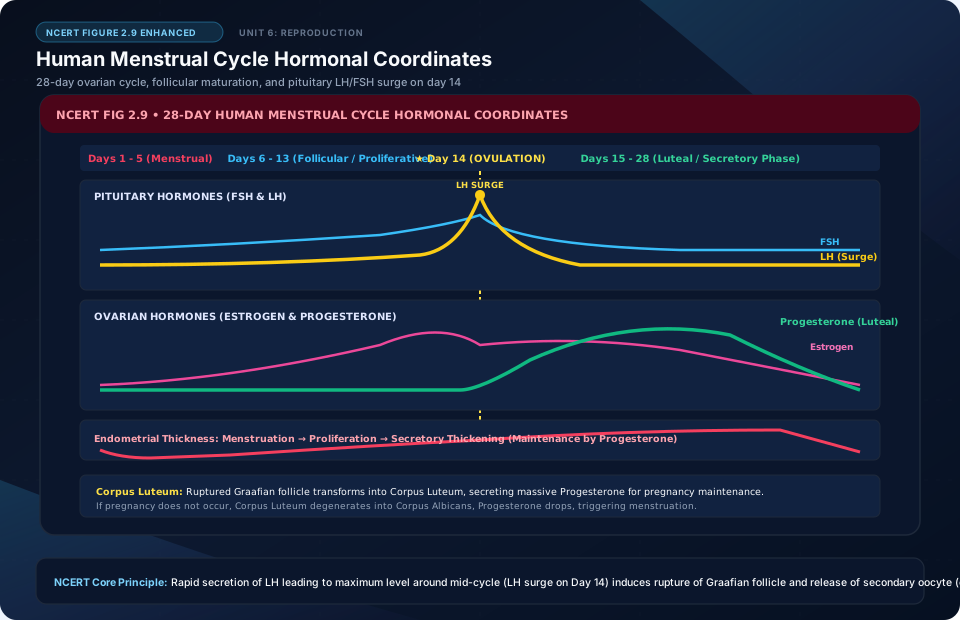
Human Menstrual Cycle Hormonal Coordinates
🚨 Examiner Traps & Common Misconceptions
✅ Reality: Oogenesis begins during EMBRYONIC fetal life! A female baby is born with all the primary oocytes she will ever have, arrested in Diplotene of Prophase I until puberty.
✅ Reality: Fertilization occurs strictly at the ampullary-isthmic junction of the Fallopian tube (oviduct)! The fertilized blastocyst then travels to implant into the uterine endometrium.
💡 Memory Hack & Mnemonic
Sperm Pathway: ‘SEVEN UP’ (Seminiferous tubules -> Epididymis -> Vas deferens -> Ejaculatory duct -> Nothing -> Urethra -> Penis).
⭐ 10/10 Active Recall Self-Check: Chapter 2
Test your mastery before closing this lecture note. Can you answer these without looking back?
- What are the core experimental proofs or physiological steps that define Human Reproduction in the NCERT syllabus?
- How does the molecular mechanism or ecological interaction covered here operate under changing conditions?
- What are the high-frequency examiner traps or tricky numerical ratios associated with this chapter?
- Can you formulate the primary mnemonic or memory framework for remembering the key components above?
⭐ 10/10 Active Recall Self-Check: Chapter 2
Test your mastery before closing this lecture note. Can you answer these without looking back?
- What are the core experimental proofs or physiological steps that define Human Reproduction in the NCERT syllabus?
- How does the molecular mechanism or ecological interaction covered here operate under changing conditions?
- What are the high-frequency examiner traps or tricky numerical ratios associated with this chapter?
- Can you formulate the primary mnemonic or memory framework for remembering the key components above?