
10.1 The cell cycle #
The study of The cell cycle provides fundamental principles governing cellular architecture, molecular interactions, and physiological regulation.

Plant Cell Structure Kn
10.1.1 Phases #
The cycle is the ordered set of events by which one cell becomes two genetically equivalent cells.
- G1 (gap 1). The cell grows, makes organelles, and decides whether to divide. A point late in G1, the restriction point in animal cells (Start in yeast), is the commitment. Before it, withdrawal of growth factors sends the cell into a quiescent state called G0. After it, the cell finishes the cycle even if growth factors fall. Many differentiated cells sit in G0 for years. G0 is reversible in cells such as lymphocytes and hepatocytes, and effectively permanent in cells such as neurons.
- S (synthesis). DNA is replicated once, and centrosomes are duplicated. The amount of DNA goes from the diploid 2C value to 4C.
- G2 (gap 2). The cell confirms that replication is complete and repairs remaining DNA damage before entering mitosis. Centrosomes mature.
- M (mitosis and cytokinesis). Chromosomes condense, segregate, and the cytoplasm splits. M is short relative to interphase in most somatic cells: about an hour, against many hours of interphase.
The whole cycle in a rapidly dividing human cell in culture is roughly 24 hours, with G1 the most variable phase. Early embryonic cycles, as in the frog egg, run S and M without growth, which is how a large egg is subdivided.
10.1.2 What must be true before division #
Three conditions are non-negotiable, and the checkpoints of Chapter 11 enforce them.
1. DNA is fully replicated, and replicated only once.
2. Damage that would be inherited is repaired, or the cycle waits.
3. Every chromosome is attached to both poles before the cohesin that holds sister chromatids is cut.
A cycle that skips any of these produces daughters with the wrong amount of DNA, which is the usual route into cell death or into the genomic instability of a tumour.
10.2 Mitosis #
The study of Mitosis provides fundamental principles governing cellular architecture, molecular interactions, and physiological regulation.

Anaphase During Mitosis
10.2.1 The chromosome and the spindle #
Before mitosis, each chromosome has been duplicated into two sister chromatids, held together along their length and especially at the centromere by the protein complex cohesin. The centromere assembles the kinetochore, the machine that binds microtubules. Condensin, a relative of cohesin, compacts the chromatids as mitosis begins.
The spindle is a microtubule apparatus with two poles, each based on a centrosome in animal cells. Three classes of microtubule do the work.
- Kinetochore microtubules attach to chromatids.
- Interpolar microtubules overlap in the middle and are slid apart by plus-end kinesins, which pushes the poles away from each other.
- Astral microtubules radiate toward the cortex and help position the spindle.
10.2.2 Stages #
Stages are a convenience for a continuous process. The useful markers are the state of the envelope, the attachment of chromosomes, and the cohesion of sisters.
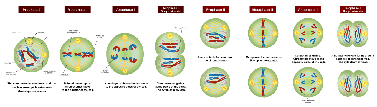
Meiosis Stages
Prophase. Chromosomes condense and become visible. Centrosomes move apart, nucleating the two asters. In the cytoplasm the spindle is assembling.
Prometaphase. The nuclear envelope breaks down in open mitosis (animals, plants). Lamins are phosphorylated, the lamina disassembles, and nuclear-pore proteins disperse. Microtubules can now reach the kinetochores. Attachment is initially unstable. A chromosome may attach to one pole only (monotelic), or both sisters may attach to the same pole (syntelic). These errors are corrected because incorrect attachments do not generate tension, and the spindle-assembly checkpoint (Chapter 11) withholds the signal to proceed. The correct, stable attachment is bi-orientation: sisters attached to opposite poles, under tension.
Metaphase. All chromosomes are aligned at the metaphase plate, bi-oriented. The cell waits here until the last kinetochore is attached. Congression to the plate is an active process of motors and of microtubule growth and shrinkage, not a passive lining-up.
Anaphase. Cohesin is cleaved (the mechanism is in Chapter 11). Two movements follow.
- Anaphase A. Chromatids move toward the poles as kinetochore microtubules shorten, largely by depolymerisation at the kinetochore and the pole, with the chromatid staying attached.
- Anaphase B. Poles move apart as interpolar microtubules slide and astral microtubules pull.
Telophase. Chromatids, now daughter chromosomes, arrive at the poles and decondense. The nuclear envelope reassembles around each set from ER membranes, lamins are dephosphorylated, and nucleoli return. The spindle disassembles.
10.2.3 Cytokinesis #
In animal cells a contractile ring of actin and myosin II assembles at the cortex in the plane of the former metaphase plate, positioned by signals from the spindle midzone. The ring constricts, making a cleavage furrow, until the connection is a midbody that is cut. The two daughters each receive a nucleus, roughly half the cytoplasm, and a share of organelles. Mitochondria and the ER are actively partitioned, not left to chance.

Mitotic Cytokinesis
In plant cells a contractile ring cannot work against a wall. The phragmoplast, a cylinder of microtubules and actin, guides Golgi-derived vesicles to the midplane. The vesicles fuse to form the cell plate, which grows outward until it joins the existing wall and splits the cytoplasm. The new wall then differentiates.
Closed mitosis, used by many fungi, keeps the envelope intact and builds the spindle inside the nucleus. The logic of bi-orientation and cohesin cleavage is the same.
10.3 Meiosis #
The study of Meiosis provides fundamental principles governing cellular architecture, molecular interactions, and physiological regulation.

Meiosis Ii Maternal Origin
10.3.1 Purpose #
Mitosis keeps the chromosome number constant. Sexual reproduction would double it at every fertilisation unless a prior division halved it. Meiosis produces haploid gametes (or haploid spores) and, while doing so, generates new combinations of alleles. Two events create the new combinations: crossing over between homologous chromosomes, and the independent orientation of different chromosome pairs.
DNA is replicated once, in the preceding S phase. Two divisions follow, meiosis I and meiosis II, with no replication between them. The products have half the DNA of the starting germ cell and are genetically non-identical.
10.3.2 Meiosis I, the reductional division #
Homologous chromosomes, one from each parent, pair along their length. This pairing, synapsis, is the event mitosis does not have. It is held by the synaptonemal complex. Crossing over occurs, with the exchange of corresponding segments between non-sister chromatids. The physical joins that result are chiasmata, and they become visible as the synaptonemal complex disassembles. Chiasmata, together with remaining cohesin along the arms, hold the homologues together so that they can be bi-oriented as a pair.

Meiosis Ii Maternal Origin (Recombinated)
Prophase I is long and is subdivided because the structures appear in order.
| Stage | What is seen |
|---|---|
| Leptotene | Chromosomes condense; homology search begins |
| Zygotene | Synapsis starts; the synaptonemal complex assembles |
| Pachytene | Synapsis is complete; crossing over occurs |
| Diplotene | The synaptonemal complex disassembles; chiasmata hold homologues together |
| Diakinesis | Chromosomes shorten further; the envelope breaks down as the spindle forms |
At metaphase I, homologues (each still two chromatids) line up as bivalents. At anaphase I, homologues separate. Sister chromatids stay together because cohesin at the centromere is protected during meiosis I and is not cleaved until meiosis II. This protection, by the protein shugoshin, is the reason meiosis I reduces the chromosome number and meiosis II then separates sisters as mitosis would.

Of Meiosis
10.3.3 Meiosis II and the genetic consequences #
Meiosis II resembles mitosis, starting from a haploid set of chromosomes that are still duplicated. The spindle attaches, protected centromeric cohesin is cleaved, and sisters separate. Four haploid nuclei result. In spermatogenesis all four become gametes. In oogenesis of many animals the cytoplasm is divided unequally, producing one egg and small polar bodies, so that the egg keeps the stores it will need after fertilisation.

Meiosis Ii Paternal Origin
Consequences to be able to state:
- Chromosome number is halved, so fertilisation restores the diploid number.
- Crossing over produces recombinant chromatids, mixing alleles along a chromosome.
- Independent assortment of different bivalents produces 2ⁿ combinations of parental chromosomes, where n is the haploid number, before counting cross-overs. In humans n = 23, so more than 8 million assortments are possible before recombination multiplies the number further.
- Errors in segregation, nondisjunction, produce gametes with an extra or a missing chromosome. Fertilisation then yields aneuploidy, for example trisomy 21. The risk rises with maternal age, in part because meiosis I in human oocytes begins before birth and arrests in diplotene for decades, and cohesin ages.

Meiosis Overview
10.4 Key terms, summary, and self-check #
Key terms. G1; S; G2; G0; restriction point; sister chromatid; cohesin; kinetochore; bi-orientation; anaphase A; anaphase B; contractile ring; cell plate; synapsis; chiasma; shugoshin; independent assortment; nondisjunction.
Summary. The cycle is G1, S, G2, and M, with an optional G0. Mitosis attaches sister chromatids to opposite poles and then splits them, followed by a contractile ring or a cell plate. Meiosis pairs homologues, recombines them, separates them at meiosis I, and separates sisters at meiosis II, producing haploid recombinant cells.
Self-check.
1. A cell has 4C DNA and condensed chromosomes aligned in one plane, with sisters attached to opposite poles. Which stage is this, and what molecular event lets it leave that stage?
2. Why does anaphase A not require the microtubules to slide, whereas anaphase B does?
3. How does a plant cell divide its cytoplasm without a contractile ring?
4. At which division do homologous chromosomes separate, and what still holds the sister chromatids together at that moment?
5. Why does a single round of premeiotic DNA replication suffice for two meiotic divisions?
—
—
—