
5.1 What are Lipids? Fatty Acid Structure & Saturation Dynamics #
Lipids represent a chemically diverse class of biological molecules unified by a single physical characteristic: insolubility in water and high solubility in nonpolar organic solvents. Unlike proteins, nucleic acids, and polysaccharides, lipids are not covalent polymers; rather, their macroscopic cellular structures (such as membranes, micelles, and liposomes) assemble spontaneously via non-covalent hydrophobic interactions driven by the liberation of ordered water cages (Δ S > 0).
Biological membranes are the defining boundaries of life. They establish the physical perimeter of the cell (plasma membrane) and compartmentalize specialized biochemical processes into intracellular organelles (mitochondria, nuclei, lysosomes, chloroplasts). Membranes are not rigid walls; they are two-dimensional fluid liquid crystals packed with proteins that carry out transport, signal transduction, and bioenergetic energy transduction.
Fatty acids are carboxylic acids with aliphatic hydrocarbon side chains ranging from 4 to 36 carbons. In biological systems, almost all fatty acids possess an even number of carbons (typically 12 to 24) because they are synthesized through the sequential addition of two-carbon acetyl-CoA units.
5.1.1 Saturated vs. Unsaturated Fatty Acids #
- Saturated Fatty Acids: Contain no carbon-carbon double bonds. The hydrocarbon chain is fully extended in the most stable staggered anti conformation, allowing saturated chains to pack tightly into crystalline arrays with extensive van der Waals contacts. Consequently, saturated fatty acids have high melting temperatures and are solid at room temperature (e.g., Palmitic acid 16:0, Tm = 63.1°C; Stearic acid 18:0, Tm = 69.6°C).
- Unsaturated Fatty Acids: Contain one or more double bonds. In virtually all naturally occurring biological unsaturated fatty acids, the double bonds are in the cis configuration. A cis double bond introduces a rigid 30° kink into the hydrocarbon chain. This kink prevents tight crystalline packing, increases intermolecular spacing, and drastically lowers the melting temperature (e.g., Oleic acid 18:1Δ 9, Tm = 13.4°C; Linoleic acid 18:2Δ 9,12, Tm = -5.0°C).
Saturated (Stearate):   ──────/──────/──────/──────/─── (Straight rod, packs tightly)
Cis-Unsaturated (Oleate):──────/──────/╭──                (30° kink, disrupts packing)
                                        ╰──────/───
—
5.2 Storage Lipids: Triacylglycerols #
Triacylglycerols (triglycerides) are composed of three fatty acids esterified to a single molecule of glycerol. They are entirely nonpolar and hydrophobic.
5.2.1 Why are Fats the Ultimate Energy Reserve? #
Triacylglycerols stored in adipocyte lipid droplets serve as the primary long-term energy reserve in animals, vastly outperforming glycogen for two biophysical reasons:
1. Higher Oxidation State (Reduced Carbons): The carbon atoms in fatty acids are almost entirely reduced hydrocarbons (-CH2-), whereas the carbons in carbohydrates are already partially oxidized (-CHOH-). The complete aerobic oxidation of 1 g of fat yields 37 kJ (9 kcal), compared to only 17 kJ (4 kcal) per gram of dry carbohydrate or protein.
2. Anhydrous Storage: Glycogen is highly polar and binds approximately 2 g of water per gram of glycogen. Triacylglycerols are completely hydrophobic and are stored in an anhydrous form. To store the energy equivalent of 15 kg of body fat, a human would have to carry over 60 kg of hydrated glycogen!
—
5.3 Membrane Lipids: Architecture and Classes #
Membranes are constructed from amphipathic lipids possessing a hydrophilic polar head group and hydrophobic hydrocarbon tails.
5.3.1 Glycerophospholipids (Phosphoglycerides) #
Consist of a glycerol-3-phosphate backbone esterified to two fatty acids at C-1 and C-2, with a polar head group attached to the phosphate at C-3:
- C-1 fatty acid is typically saturated (16:0 or 18:0).
- C-2 fatty acid is typically unsaturated (18:1 or 20:4).
- Common head groups include: Choline (Phosphatidylcholine, PC), Ethanolamine (Phosphatidylethanolamine, PE), Serine (Phosphatidylserine, PS, carrying a net negative charge), and Inositol (Phosphatidylinositol, PI, involved in IP3/DAG cell signaling).
The complete chemical composition and atomic topology of a representative phospholipid are detailed in Figure 5.1.

Phospholipid Chemical Structure
5.3.2 Sphingolipids #
Built on the amino alcohol sphingosine (an 18-carbon unsaturated amino alcohol) rather than glycerol. A fatty acid is linked via an amide bond to the C-2 amino group, forming a ceramide.
- Sphingomyelin: Ceramide attached to a phosphocholine head group; constitutes a major structural component of the myelin sheath insulating nerve axons.
- Glycosphingolipids (Cerebrosides and Gangliosides): Ceramide attached to one or more sugar residues (lacking phosphate). Gangliosides contain complex branched oligosaccharides terminating in negatively charged sialic acid (NANA) residues; they dominate the outer surface of neuronal membranes and act as receptors for bacterial toxins (e.g., cholera toxin).
5.3.3 Cholesterol and Sterols #
Cholesterol is an amphipathic 27-carbon lipid consisting of a rigid, planar steroid nucleus (four fused rings: three 6-membered cyclohexane rings and one 5-membered cyclopentane ring in a trans conformation), a nonpolar hydrocarbon tail at C-17, and a single polar hydroxyl group (-OH) at C-3.
The atomic structure and steroid ring system of cholesterol are depicted in Figure 5.2.
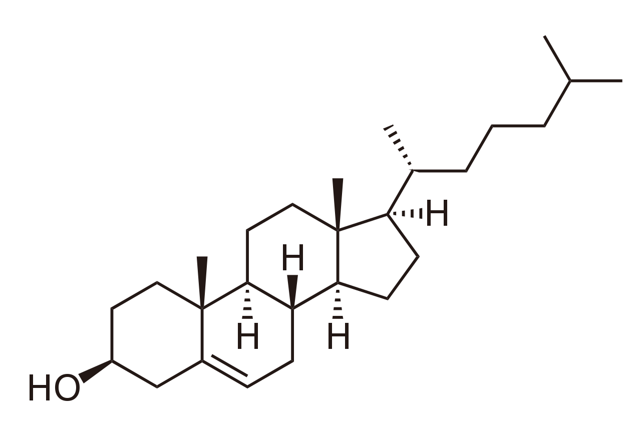
Cholesterol
—
5.4 Supramolecular Self-Assembly: Micelles, Liposomes, and Bilayers #
In aqueous solution, amphipathic lipids spontaneously assemble into organized macroscopic structures to minimize the contact between hydrophobic tails and water:
- Micelles: Formed by single-tailed lipids with a conical geometry (e.g., free fatty acids, SDS detergents). The cross-sectional area of the head group is larger than that of the single acyl chain.
- Lipid Bilayers: Formed by two-tailed phospholipids and sphingolipids with a cylindrical geometry. Two parallel monolayers (leaflets) form a continuous planar sheet with an interior hydrophobic core (~ 30 AA thick) flanked by polar head groups (~ 15 AA each, total thickness ~ 50–60 AA).
- Liposomes (Vesicles): A planar bilayer folds back upon itself to enclose an aqueous cavity, eliminating all exposed hydrophobic edges. Liposomes are used clinically as targeted nanoparticle delivery systems for hydrophobic chemotherapeutics and mRNA vaccines.
The three primary supramolecular structures formed by phospholipids in aqueous solutions are compared in Figure 5.3.

Phospholipids Aqueous Solution Structures
—
5.5 Membrane Dynamics & The Fluid Mosaic Model #
In 1972, S. J. Singer and Garth L. Nicolson proposed the Fluid Mosaic Model: membranes are two-dimensional liquid crystalline solutions of oriented lipids and globular integral proteins.
5.5.1 Membrane Fluidity and Phase Transitions #
A lipid bilayer undergoes a characteristic phase transition from a rigid, immobilized gel phase (paracrystalline state) to a dynamic, disordered liquid-disordered fluid phase at a specific transition temperature (Tm).
Fluidity is regulated by:
1. Fatty Acid Chain Length & Unsaturation: Shorter chains and more cis double bonds depress Tm, keeping membranes fluid at lower temperatures (Homeoviscous Adaptation in cold-tolerant fish and bacteria).
2. Cholesterol as a Bidirectional Fluidity Buffer:
- At High Temperatures (above Tm): The rigid steroid ring system intercalates between acyl chains, restricting their rotational flailing, decreasing membrane fluidity, and preventing the membrane from becoming leaky.
- At Low Temperatures (below Tm): Cholesterol’s bulky ring prevents saturated hydrocarbon chains from packing closely together and crystalizing, preventing the membrane from freezing solid.
5.5.2 Transbilayer Asymmetry & Flippases #
Membranes exhibit stark compositional asymmetry between the outer (exofacial) and inner (cytoplasmic) leaflets:
- Outer Leaflet: Rich in Phosphatidylcholine (PC), Sphingomyelin, and Glycolipids.
- Inner Leaflet: Rich in Phosphatidylethanolamine (PE), Phosphatidylinositol (PI), and Phosphatidylserine (PS).
Lateral diffusion within a leaflet is extremely rapid (D ~ 10-8 cm2/s; a lipid travels the length of a bacterium in 1 second). In contrast, spontaneous transverse diffusion (“flip-flop”) across the hydrophobic core is extraordinarily slow (t1/2 ~ days to weeks) because the polar head group must traverse the anhydrous core.
To establish and maintain asymmetry, cells express ATP-dependent transport enzymes:
- Flippase (P-type ATPase): Moves PS and PE from the outer leaflet to the inner leaflet against a concentration gradient, consuming ATP.
- Floppase (ABC transporter): Moves phospholipids from the inner to the outer leaflet, consuming ATP.
- Scramblase: A bidirectional, Ca2+-activated transporter that dissipates lipid asymmetry down concentration gradients without consuming ATP.
> Apoptotic Signaling Rule: When an aged or damaged cell enters apoptosis, intracellular Ca2+ surges and scramblase is activated, while flippase is cleaved by caspases. Phosphatidylserine (PS) exposes on the outer leaflet, acting as an “eat-me” signal that binds scavenger receptors on macrophages, triggering phagocytosis without inducing an inflammatory response!
—
5.6 Curiosity Spark & Clinical Insights #
—
5.7 Key Takeaways & High-Yield Summary #
- Thermodynamic driver: Lipid self-assembly into bilayers is driven by the hydrophobic effect (positive water entropy Δ S > 0), not by covalent bonding.
- Saturation and fluidity: Saturated fatty acids pack into solid crystalline arrays; cis double bonds introduce 30° kinks that disrupt packing, depressing Tm.
- Energy storage superiority: Triacylglycerols yield 37 kJ/g and store anhydrously, whereas glycogen yields 17 kJ/g and binds 2 g water per gram.
- Cholesterol buffering: Restricts high-temperature fluidity while preventing low-temperature crystalline freezing.
- Phosphatidylserine asymmetry: Maintained on the inner leaflet by ATP-dependent flippases; externalization to the outer leaflet signals apoptotic phagocytosis.
—
5.8 Review & Self-Assessment Questions #
1. Explain why deep-sea hydrothermal bacteria and psychrophilic polar ocean bacteria exhibit vastly different membrane lipid compositions.
- Answer: Psychrophilic bacteria living at 0–4°C incorporate high percentages of short-chain and polyunsaturated cis-fatty acids to prevent membrane freezing. Thermophilic bacteria at >80°C incorporate long-chain saturated fatty acids and ether-linked branched isoprenoids to prevent membrane dissolution and thermal leakage.
2. What occurs when red blood cells are treated with a purified flippase inhibitor?
- Answer: Over time, spontaneous or scramblase-mediated leakage causes Phosphatidylserine (PS) to accumulate on the outer leaflet. Without active flippase to pump PS back to the inner leaflet, externalized PS will be recognized by splenic macrophages, leading to premature hemolytic clearance of the red blood cells.
—