
2.1 What is an Amino Acid? Chemical Architecture & Stereochemistry #
An amino acid is a bifunctional organic molecule containing both a basic amino group (-NH2) and an acidic carboxylic acid group (-COOH). In all biological proteins, both functional groups are covalently bonded to the exact same central carbon atom—designated the alpha-carbon (C_α). Because both groups share the α-carbon, these monomers are specifically termed α-amino acids.
Every standard α-amino acid possesses four distinct chemical groups bonded to the tetrahedral C_α carbon:
1. An α-carboxyl group (-COOH).
2. An α-amino group (-NH2).
3. A hydrogen atom (-H).
4. A distinct, variable chemical side chain designated the R-group.
The fundamental molecular architecture of a generic α-amino acid is illustrated in Figure 2.1, showing the spatial orientation of the chiral alpha carbon and its surrounding functional groups.
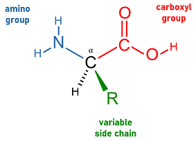
Amino Acid Generic Structure
2.1.1 Chirality and the L-Stereoisomer Rule #
Because four chemically distinct substituents occupy the tetrahedral vertices of the C_α carbon, the α-carbon constitutes a chiral stereocenter (asymmetric carbon). Consequently, amino acids exist as two non-superimposable mirror-image enantiomers: D-amino acids and L-amino acids.
- Glycine is the sole exception: Glycine’s R-group is a single hydrogen atom. Having two identical hydrogen atoms bonded to C_α, glycine is achiral and optically inactive.
- The L-Enantiomer Rule in Biology: Ribosomal protein synthesis across all domains of life (Archaea, Bacteria, Eukarya) uses exclusively L-α-amino acids. (In Fischer projections, the α-amino group projects to the left when the carboxyl group is drawn at the top).
- D-amino acids exist in nature only in specialized non-ribosomal bacterial peptides (such as the peptidoglycan cell wall) and peptide antibiotics (valinomycin, gramicidin).
- Why homochirality matters: Homochirality is an absolute physical necessity for structural biology. A polypeptide chain constructed from a racemic mixture of D- and L-amino acids cannot form stable secondary structures such as right-handed α-helices or β-sheets because the alternating orientations of side chains cause catastrophic steric clash.
—
2.2 Acid-Base Properties: Zwitterions, Titration Curves & Isoelectric Point (pI) #
In an aqueous solution at physiological pH (7.4), amino acids never exist as un-ionized molecules (-NH2 and -COOH). Instead, the carboxylic acid (pKa1 ≈ 2.0) loses its proton to become a carboxylate ion (-COO–), while the basic amino group (pKa2 ≈ 9.5) accepts a proton to become an ammonium ion (-NH3+).
Molecules carrying simultaneous positive and negative charges are called zwitterions (dipolar ions). A zwitterion is electrically neutral overall, but carries localized charges.
      At pH 1.0 (Cationic)           At pH 7.4 (Zwitterion)          At pH 12.0 (Anionic)
              H                               H                               H
              │                               │                               │
       H3N+ ─ Cα ─ COOH               H3N+ ─ Cα ─ COO-                H2N ─ Cα ─ COO-
              │                               │                               │
              R                               R                               R
        (Net Charge = +1)               (Net Charge = 0)               (Net Charge = -1)
The experimental titration curves of the 20 amino acids categorized by their side-chain ionization properties are presented in Figure 2.2, showing the inflection points corresponding to pKa1, pKa2, and side-chain pKaR.

Titration Curves Of 20 Amino Acids Organized By Side Chain
2.2.1 Calculating the Isoelectric Point (pI) #
The isoelectric point (pI) is the precise pH at which the net electrical charge of an amino acid (or protein) is exactly zero.
1. For Diprotic Amino Acids (Non-ionizable side chains, e.g., Alanine):
pI = frac{pKa1 (α-COOH) + pKa2 (α-NH3+)}{2} = (2.34 + 9.69 / 2) = 6.01
2. For Acidic Amino Acids (Carboxyl side chains: Aspartate, Glutamate): The net-neutral species exists between the deprotonation of the α-carboxyl and the R-group carboxyl:
pI = frac{pKa1 (α-COOH) + pKaR (side-chain carboxyl)}{2} = (1.88 + 3.65 / 2) = 2.77 quad (Aspartate)
3. For Basic Amino Acids (Cationic side chains: Lysine, Arginine, Histidine): The net-neutral species exists between the deprotonation of the α-amino and the side-chain amino:
pI = frac{pKa2 (α-NH3+) + pKaR (varepsilon-amino)}{2} = (8.95 + 10.53 / 2) = 9.74 quad (Lysine)
At any pH < pI, the amino acid carries a net positive charge and migrates toward the cathode during electrophoresis. At any pH > pI, it carries a net negative charge and migrates toward the anode.
—
2.3 The Twenty Standard Amino Acids: Chemical Classification #
The variable side chain (R-group) dictates the unique solubility, chemical reactivity, and structural role of each amino acid. The standard twenty amino acids are categorized into five distinct chemical families:
1. Nonpolar, Aliphatic R-Groups (7 Residues) #
- Glycine (Gly, G): Single hydrogen atom as side chain; achiral, smallest residue. Imparts immense conformational flexibility to loop regions, reverse turns, and collagen triple helices.
- Alanine (Ala, A): Has a small methyl group (-CH3); universally accommodated in α-helices.
- Valine (Val, V), Leucine (Leu, L), Isoleucine (Ile, I): Branched-chain aliphatic hydrocarbons. Form the densely packed hydrophobic cores of globular proteins via van der Waals interactions.
- Methionine (Met, M): Nonpolar thioether (-CH2-CH2-S-CH3). Universal initiating residue in eukaryotic and bacterial translation.
- Proline (Pro, P): Unique cyclic secondary amino acid (imino acid). Its side chain loops back to form a five-membered ring with its own α-amino nitrogen. This rigid ring locks the backbone φ angle near -60°, introducing sharp kinks and acting as a powerful α-helix breaker.
2. Aromatic R-Groups (3 Residues) #
- Phenylalanine (Phe, F): Purely hydrophobic nonpolar benzyl ring.
- Tyrosine (Tyr, Y): Phenol group with a weakly ionizable hydroxyl (pKa ≈ 10.1); participates in hydrogen bonding and is a primary target of receptor tyrosine kinases.
- Tryptophan (Trp, W): Bulky bicyclic indole ring.
> Ultraviolet Spectroscopy Rule: Tyrosine and Tryptophan account for the characteristic ultraviolet absorption of proteins at λ = 280 nm, enabling rapid concentration measurements via the Beer-Lambert law (A = varepsilon c l).
3. Polar, Uncharged R-Groups (5 Residues) #
- Serine (Ser, S) & Threonine (Thr, T): Possess polar hydroxyl groups (-OH) that form hydrogen bonds and serve as primary phosphorylation sites for regulatory kinases.
- Cysteine (Cys, C): Possesses a reactive sulfhydryl (thiol, -SH) group (pKa ≈ 8.3). Under oxidizing conditions (such as the endoplasmic reticulum lumen or extracellular space), two cysteines oxidize to form a covalent disulfide bond (cystine, -S-S-), locking extracellular proteins (e.g., insulin, antibodies) into stable folds.
- Asparagine (Asn, N) & Glutamine (Gln, Q): Amides of aspartate and glutamate; polar, neutral hydrogen bonders. Asn is the site of N-linked glycosylation.
4. Positively Charged (Basic) R-Groups (3 Residues) #
- Lysine (Lys, K): Features an aliphatic chain ending in a primary varepsilon-amino group (pKa ≈ 10.5). Positive at physiological pH; target of ubiquitination, acetylation, and methylation.
- Arginine (Arg, R): Features a planar guanidinium group with a high pKa ≈ 12.5. The positive charge is extensively delocalized across all three nitrogen atoms by resonance.
- Histidine (His, H): Possesses an aromatic imidazole ring with a side-chain pKa ≈ 6.0. Because its pKa is near physiological neutrality, histidine shifts easily between protonated and neutral states, making it the premier general acid-base catalytic proton shuttle in enzyme active sites (e.g., chymotrypsin, ribonuclease).
5. Negatively Charged (Acidic) R-Groups (2 Residues) #
- Aspartate (Asp, D) & Glutamate (Glu, E): Possess β- and γ-carboxyl groups with pKa ≈ 3.7–4.3. Fully deprotonated (-COO–) and negatively charged at pH 7.4. Essential for forming ionic salt bridges and coordinating catalytic divalent cations (Mg2+, Ca2+).
| Amino Acid | 3-Letter | 1-Letter | Side-Chain Class | Side-Chain pKa | Isoelectric Point (pI) |
|---|---|---|---|---|---|
| Glycine | Gly | G | Nonpolar aliphatic | — | 5.97 |
| Alanine | Ala | A | Nonpolar aliphatic | — | 6.01 |
| Valine | Val | V | Nonpolar branched | — | 5.97 |
| Leucine | Leu | L | Nonpolar branched | — | 5.98 |
| Isoleucine | Ile | I | Nonpolar branched | — | 6.02 |
| Methionine | Met | M | Nonpolar thioether | — | 5.74 |
| Proline | Pro | P | Nonpolar cyclic imino | — | 6.48 |
| Phenylalanine | Phe | F | Aromatic | — | 5.48 |
| Tyrosine | Tyr | Y | Aromatic / Polar | 10.07 | 5.66 |
| Tryptophan | Trp | W | Aromatic | — | 5.89 |
| Serine | Ser | S | Polar uncharged | — | 5.68 |
| Threonine | Thr | T | Polar uncharged | — | 5.60 |
| Cysteine | Cys | C | Polar sulfhydryl | 8.18 | 5.07 |
| Asparagine | Asn | N | Polar amide | — | 5.41 |
| Glutamine | Gln | Q | Polar amide | — | 5.65 |
| Lysine | Lys | K | Positively charged (basic) | 10.53 | 9.74 |
| Arginine | Arg | R | Positively charged (basic) | 12.48 | 10.76 |
| Histidine | His | H | Positively charged (imidazole) | 6.00 | 7.59 |
| Aspartate | Asp | D | Negatively charged (acidic) | 3.65 | 2.77 |
| Glutamate | Glu | E | Negatively charged (acidic) | 4.25 | 3.22 |
—
2.4 The Peptide Bond: Planarity, Resonance & Dihedral Constraints #
Amino acids polymerize via a condensation (dehydration) reaction between the α-carboxyl group of one amino acid and the α-amino group of another, eliminating water to form a peptide bond (amide linkage).
The stereochemical condensation mechanism and the partial double-bond character of the resulting peptide bond are illustrated in Figure 2.3.

Peptide Bond Formation
2.4.1 Partial Double-Bond Character & Planarity #
Linus Pauling and Robert Corey demonstrated that the peptide C-N bond exhibits an unusually short length (1.33 AA), significantly shorter than a single C-N bond (1.45 AA) and approaching a C=N double bond (1.25 AA).
This occurs because the nitrogen lone pair delocalizes into the carbonyl π-system, giving the peptide bond ~ 40% double-bond character:
O=C-N-H longleftrightarrow –O-C=N+-H
#### Biophysical Consequences:
1. Planarity: Rotation around the peptide C-N bond is strictly forbidden at physiological temperatures (activation barrier ~ 80 kJ/mol). The six atoms of the peptide unit (C_α – C(=O) – N(-H) – C_α) are locked in a single rigid plane.
2. Trans Preference: Nearly all peptide bonds adopt the trans configuration (adjacent C_α atoms on opposite sides of the C-N bond), which avoids steric clash between adjacent side chains. Trans is favored over cis by 1000:1, except before Proline, where ~ 6% of X-Pro bonds exist as cis (isomerized by peptidyl prolyl cis-trans isomerases).
3. Dihedral Angles (φ and ψ): The conformational freedom of the polypeptide backbone is restricted to rotations around two single bonds flanking C_α:
- φ (phi): Angle around the N-C_α bond.
- ψ (psi): Angle around the C_α-C (carbonyl) bond.
The Ramachandran Plot maps all sterically permissible (φ, ψ) angle pairs. Because non-bonded atoms collide at most angles, over 75% of conformational space is forbidden! The allowed islands correspond to regular secondary structures: α-helices (lower left) and β-sheets (upper left).
—
2.5 Hierarchical Levels of Protein Structure #
Proteins fold into four hierarchical levels of structural complexity, proceeding from linear covalent sequences to multimeric complexes.
Figure 2.4 illustrates this structural hierarchy across primary, secondary, tertiary, and quaternary levels.

Main Protein Structure Levels
2.5.1 Primary Structure #
The linear sequence of amino acids joined by covalent peptide bonds, read from the N-terminus to the C-terminus, along with the positions of covalent disulfide bridges. Primary structure contains all information needed to direct spontaneous folding into the native tertiary state (Anfinsen’s Dogma).
2.5.2 Secondary Structure: α-Helices, β-Sheets, and Turns #
Regular, local backbone conformations stabilized exclusively by hydrogen bonds between backbone carbonyl oxygens (C=O) and backbone amide nitrogens (N-H):
- The α-Helix: A right-handed helical rod with 3.6 residues per turn and an axial pitch of 0.54 nm (5.4 AA). Every backbone C=O of residue i forms a direct hydrogen bond with the N-H of residue i+4. All hydrogen bonds run parallel to the helix axis. Side chains project outward. The helix carries a macroscopic dipole moment, partial positive at the N-terminus and partial negative at the C-terminus. Proline and Glycine act as helix breakers.
- β-Pleated Sheets: Formed by extended polypeptide strands (β-strands). Hydrogen bonds form between adjacent strands:
- Antiparallel β-sheets: Strands run in opposite directions; inter-strand hydrogen bonds are linear (180°) and stronger.
- Parallel β-sheets: Strands run in the same direction; hydrogen bonds are distorted and slightly weaker.
- β-Turns (Reverse Turns): Compact 4-residue hairpins that reverse chain direction by 180°. Stabilized by an internal hydrogen bond between residue i and i+3. Typically contain Glycine (flexibility) and Proline (cis conformation).
2.5.3 Tertiary Structure: The Three-Dimensional Fold #
The overall 3D folding of a single polypeptide chain, bringing sequentially distant residues into close proximity. Tertiary folding is driven primarily by:
1. The Hydrophobic Collapse: Burying nonpolar residues (Leu, Ile, Val, Phe) into the core, liberating ordered water cages and providing a large entropic boost (Δ Swater > 0).
2. Van der Waals packing: Dense core packing.
3. Electrostatic Salt Bridges: Ionic pairing between oppositely charged side chains (e.g., Asp– ·s Lys+).
4. Hydrogen bonding and Disulfide cross-links.
2.5.4 Quaternary Structure: Multimeric Cooperativity #
The spatial association of two or more independent polypeptide subunits into an oligomeric functional complex (e.g., hemoglobin α2β2). Quaternary structure enables allosteric regulation and cooperativity.
—
2.6 Allosteric Cooperativity: The Hemoglobin Model #
Hemoglobin (Hb) is the paradigm for understanding allostery and cooperativity in quaternary proteins.
- Myoglobin: Monomeric (153 residues); hyperbolic oxygen-binding curve (P50 ≈ 2.8 mmHg); serves as an oxygen storage reservoir in muscle.
- Hemoglobin (HbA): Heterotetramer (α2β2); sigmoidal (S-shaped) oxygen-binding curve (P50 ≈ 26 mmHg). It binds oxygen with >98% saturation in the lungs (PO2 ≈ 100 mmHg) and unloads >66% in working tissues (PO2 ≈ 20–40 mmHg).
The crystallographic structure of oxyhemoglobin (PDB: 2DN1) is illustrated in Figure 2.5, displaying the α2β2 tetrameric architecture and the central regulatory cavity.

2Dn1 Oxyhemoglobin Structure
2.6.1 The T-to-R Allosteric Switch #
Hemoglobin shifts between two quaternary states:
1. T-State (Tense State): Deoxyhemoglobin. Stabilized by 8 inter-subunit ionic salt bridges and by bound 2,3-bisphosphoglycerate (2,3-BPG). Has low oxygen affinity.
2. R-State (Relaxed State): Oxyhemoglobin. When O2 binds to the heme Fe2+, the iron atom is pulled into the porphyrin plane. This drags the proximal histidine and Helix F, triggering a 15° rotation of the α1β1 dimer relative to the α2β2 dimer. This rotation breaks salt bridges and expels 2,3-BPG, snapping remaining subunits into the high-affinity R-state.
The degree of cooperativity is quantified by the Hill Equation:
log≤ft((θ / 1 – θ)right) = nH log(PO2) – nH log(P50)
For human hemoglobin, nH ≈ 2.8–3.0, demonstrating strong positive cooperativity.
2.6.2 Heterotropic Allosteric Modulators #
Several physiological metabolites act as heterotropic inhibitors, stabilizing the T-state and shifting the oxygen dissociation curve to the right (facilitating tissue oxygen unloading):
1. The Bohr Effect (H+ Protons): Hypoxic, actively metabolizing tissues produce lactic and carbonic acids. Lower pH protonates His146 on β-subunits, forming a salt bridge with Asp94 that locks the low-affinity T-state, dumping oxygen into hypoxic tissue.
2. Carbon Dioxide (CO2): Binds to globin N-termini, forming negatively charged carbamino-hemoglobin, releasing protons and stabilizing the T-state.
3. 2,3-Bisphosphoglycerate (2,3-BPG): Carries a -5 charge and binds to a positively charged pocket inside the central cavity of the T-state. Without 2,3-BPG, hemoglobin stays in the R-state and unloads almost no oxygen. In fetal hemoglobin (HbF, α2γ2), His143 is replaced by Serine, reducing 2,3-BPG binding and shifting the curve leftward, enabling fetal blood to extract oxygen from maternal circulation.
—
2.7 Curiosity Spark & Clinical Insights #
—
2.8 Key Takeaways & High-Yield Summary #
- Universal structure: α-amino acids feature -NH2, -COOH, -H, and -R on a chiral C_α. Ribosomal translation uses exclusively L-enantiomers.
- Zwitterion equilibria: At physiological pH 7.4, amino acids carry both -COO– and -NH3+; pI is the pH of zero net charge.
- Peptide bond planarity: 40% double-bond character prevents rotation; conformational flexibility is restricted to backbone φ and ψ angles on the Ramachandran plot.
- Structural hierarchies: Primary (sequence); Secondary (α-helices with i → i+4 H-bonds, β-sheets); Tertiary (hydrophobic core collapse); Quaternary (multimer cooperativity).
- Allosteric shift: Hemoglobin rotates 15° from low-affinity T-state to high-affinity R-state. H+, CO2, and 2,3-BPG stabilize the T-state to promote peripheral oxygen unloading.
—
2.9 Review & Self-Assessment Questions #
1. Explain why a solution of Glycine cannot rotate plane-polarized light, whereas solutions of the other nineteen standard amino acids are optically active.
- Answer: Optical activity requires an asymmetric chiral center. In glycine, the α-carbon is bonded to two identical hydrogen atoms, giving the molecule a plane of symmetry and rendering it achiral and optically inactive. All other nineteen amino acids possess four chemically distinct substituents on C_α, creating a chiral center that rotates plane-polarized light.
2. Why does mutating a single residue from Glycine to Proline in an α-helix disrupt the helical structure?
- Answer: Proline lacks a free hydrogen atom on its backbone nitrogen when engaged in a peptide bond, making it incapable of donating the essential i → i+4 hydrogen bond. Furthermore, proline’s rigid five-membered pyrrolidine ring locks its φ dihedral angle at -60°, creating an unavoidable steric clash that kinks and terminates the α-helix.
3. Calculate the isoelectric point (pI) of Histidine given pKa1 (α-COOH) = 1.82, pKa2 (α-NH3+) = 9.17, and pKaR (imidazole) = 6.00.
- Answer: Histidine’s neutral zwitterion exists between the loss of a proton from the imidazole side chain (pKaR = 6.00) and the α-amino group (pKa2 = 9.17). Therefore: pI = (6.00 + 9.17) / 2 = 7.59.
—